The paper’s first author, Manya Malhotra discovered photonic origami by chance while trying to locate an invisible laser beam.

TAU Researchers Fold Glass into Microscopic 3D Optical Devices

With a new method called photonic origami, researchers can bend ultra-thin glass sheets into complex, ultra-smooth structures directly on a chip — a step toward new optical devices for data processing, sensing, and experimental physics.

“Existing 3D printers produce rough 3D structures that aren’t optically uniform and thus can’t be used for high-performance optics,” said research team leader Prof. Tal Carmon from the School of Electrical Engineering, Fleischman Faculty of Engineering, at Tel Aviv University in Israel. “Mimicking the way a pinecone’s scales bend outward to release seeds, our laser-induced technique triggers precise bending in ultra-thin glass sheets and can be used to create highly transparent, ultra-smooth 3D microphotonic devices for a variety of applications.”

In OpticaOptica Publishing Group’s journal for high-impact research, the researchers reported that the new laser-induced folding method can create 3-mm-long structures just 0.5 microns thick — about 1/200th the width of a human hair — setting a record length-to-thickness ratio of 3D structures. They also created helix shapes as well as concave and convex mirrors with surfaces so smooth — less than a nanometer of variation — that light reflects off them without distortion.

“Similar to how large 3D printers can fabricate almost any household item, photonic origami could enable a variety of tiny optical devices,” said Carmon. “For example, it can be used to generate micro-zoom lenses that could replace the five separate cameras used in most smartphones or to fabricate microphotonic components that use light instead of electricity — helping drive the shift toward faster, more efficient alternatives to traditional electronics in our computers.”

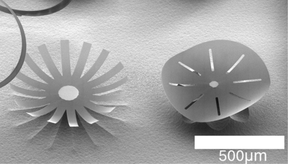
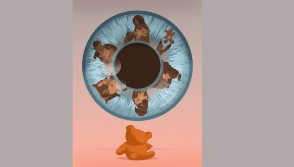
Structures made with photonic origami

Folded by accident

The new method was discovered by chance when Carmon asked graduate student Manya Malhotra to pinpoint where an invisible laser was hitting the glass by increasing the power until the spot glowed. Instead of glowing, the glass folded — revealing a simple and unexpected way to achieve glass folding. Malhotra then became the pioneering expert in photonic origami.

The glass folds because, as one side is heated with a laser, the glass liquifies and surface tension becomes stronger than gravity. As the surface tension increases, the glass is pulled into a fold precisely where the laser hits.

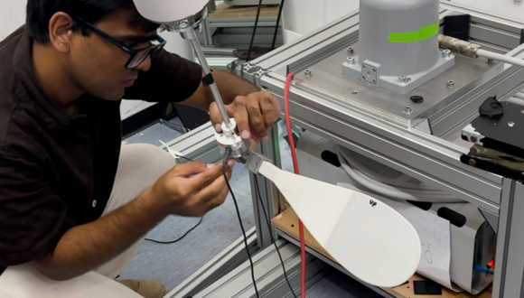
To apply this discovery, lab engineer Ronen Ben Daniel fabricated a thin layer of silica glass on a silicon chip and then shaped it into the required two-dimensional form. Before bending the glass, the researchers used etching to undercut the silicon beneath the glass sheet while leaving a small support region to hold it in place. Using CO2 laser pulses, they showed that thin glass sheets on a silicon chip could be folded in less than a millisecond, with a speed of 2 m/s and acceleration exceeding 2000 m/s2.

“It was exciting to see the folding silica under the microscope,” said Carmon. “The level of control we had over 3D microphotonic architecture came as a pleasant surprise — especially given that it was achieved with a simple setup involving just a single laser beam focused on the desired fold.”
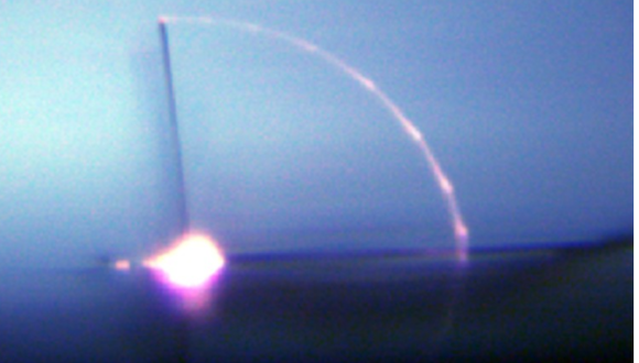
Folding glass bar

Creating microscopic structures

Using the new photonic origami approach, the researchers were able to bend sheets of glass up to 10 microns thick into shapes ranging from a 90-degree knee to helices. They were able to do this with fine control, down to 0.1 microradians.

They also used the new approach to create an extremely lightweight and precise table structure containing a concave cavity mirror, a type of mirror that focuses light. This structure was inspired by a theoretical paper by P.K. Lam from the Australian National University that proposed exploring potential deviations from Newtonian gravity at very small scales using optically levitated cavity mirrors that might be possible to fabricate using photonic origami.

To make the tiny table light enough, the researchers began with a glass sheet just 1/20 the thickness of a human hair (5 microns). They patterned the sheet much like a child’s foldable paper table toy and used their photonic origami technique to fold it into a 3D table after fabricating a concave mirror at the base of the table.

According to the researchers, this ultra-light, compact table could, in principle, be optically levitated and used to explore possible deviations from Newtonian gravity. These types of experiments could provide insights into astronomical mysteries associated with dark matter —the only area in physics where experimental observations consistently defy current theoretical predictions.

“High-performance, 3D microphotonics had not been previously demonstrated,” said Carmon. “This new technique brings silica photonics — using glass to guide and control light — into the third dimension, opening up entirely new possibilities for high-performance, integrated optical devices.”

About Optica

Optica is an open-access journal dedicated to the rapid dissemination of high-impact peer-reviewed research across the entire spectrum of optics and photonics. Published monthly by Optica Publishing Group, the Journal provides a forum for pioneering research to be swiftly accessed by the international community, whether that research is theoretical or experimental, fundamental or applied. Optica maintains a distinguished editorial board of more than 60 associate editors from around the world and is overseen by Editor-in-Chief Prem Kumar, Northwestern University, USA. For more information, visit Optica.

About Optica Publishing Group

Optica Publishing Group is a division of the society, Optica, Advancing Optics and Photonics Worldwide. It publishes the largest collection of peer-reviewed and most-cited content in optics and photonics, including 18 prestigious journals, the society’s flagship member magazine, and papers and videos from more than 835 conferences. With over 400,000 journal articles, conference papers and videos to search, discover and access, our publications portfolio represents the full range of research in the field from around the globe.

Prof. Tal Dvir, Head of the Sagol Center for Regenerative Biotechnology, Head of the Nanotechnology Center at Tel Aviv University, and Chief Scientist of the biotech company Matricelf.

Israeli Breakthrough: First Human Trial of Engineered Spinal Cord

TAU researchers begin to prepare for implantation of engineered spinal cord tissues in humans — a new development that brings fresh hope to paralyzed patients.

What if we could restore the ability to walk to people paralyzed by injury or illness?

This vision is now moving closer to reality. Three years ago, Tel Aviv University researchers succeeded in engineering a human spinal cord in the lab for the first time. Since then, progress has been rapid, with animal trials showing unprecedented success. Now, for the first time, the technology is set to be tested in human patients.

Prof. Tal Dvir, of TAU’s Sagol Center for Regenerative Biotechnology, head of TAU’s Jan Koum Center for Nanoscience and Nanotechnology, and Chief Scientist of the biotech company Matricelf, explains:
“The spinal cord is made up of nerve cells that transmit electrical signals from the brain to every part of the body. When the spinal cord is torn due to trauma — from a car accident, a fall, or a battlefield injury — this chain is broken. Think of it like an electrical cable that’s been cut: if the two parts don’t touch, the electrical signal can’t pass. The cable won’t carry electricity, and in the same way, the person can’t transmit the signal beyond the site of the injury.”

This is one of the few injuries in the human body with no natural ability to regenerate. “Neurons are cells that do not divide and do not renew themselves. They are not like skin cells, which can repair themselves after injury. They are more similar to heart cells: once damage occurs, the body cannot restore them,” notes Prof. Dvir.

Engineering a Personalized Implant

To overcome this challenge, the TAU researchers developed a fully personalized process. Blood cells are taken from the patient and reprogrammed through genetic engineering to behave like embryonic stem cells, capable of becoming any type of cell in the body.

Meanwhile, fat tissue from the same patient is used to extract substances such as collagen and sugars. These are used to produce a unique hydrogel. “The beauty of this gel is that it’s also personalized, just like the cells. We take the cells that we’ve reprogrammed into embryonic-like stem cells, place them inside the gel, and mimic the embryonic development of the spinal cord,” says Prof. Dvir.

The result is a complete three-dimensional implant. “At the end of the process, we don’t just turn the cells into motor neurons — because cells alone won’t help us — but into three-dimensional tissue: neuronal networks of the spinal cord. After about a month, we obtain a 3D implant with many neurons that transmit electrical signals. These 3D tissues are then implanted into the damaged area.”

Visualization of the next stage of the research – human spinal cord implants for treating paralysis (Photo: Sagol Center for Regenerative Biotechnology)

From Animals to Human Patients

The researchers first tested the implant in lab animals. “We showed that we can treat animals with chronic injuries. Not animals that were injured just recently, but those we allowed enough time to pass — like a person more than a year after an injury. More than 80% of the animals regained full walking ability,” Prof. Dvir explains.

Encouraged by these results, the team submitted the findings to Israel’s Ministry of Health. “About six months ago we received preliminary approval to begin compassionate-use trials with eight patients. We decided, of course, that the first patient would be Israeli. This is undoubtedly a matter of national pride. The technology was developed here in Israel, at Tel Aviv University and at Matricelf, and from the very beginning it was clear to us that the first-ever surgery would be performed in Israel, with an Israeli patient.” he says.

Looking Ahead

The first implant in a human patient is expected within about a year. For the initial trials, the team will focus on patients whose paralysis is relatively recent — within about a year of injury. “Once we prove that the treatment works — everything is open, and we’ll be able to treat any injury,” says Prof. Dvir.

Behind the initiative are key figures from both academia and industry. Prof. Dvir founded Matricelf in 2019 together with Dr. Alon Sinai, based on the revolutionary organ engineering technology developed at TAU under a licensing agreement through Ramot, the University’s technology transfer company. The company’s CEO is Gil Hakim, while the scientific development is led by Dr. Tamar Harel-Adar and her team.

“They managed to get us to the stage of regulatory approvals so quickly — and that’s amazing,” says Prof. Dvir.

Gil Hakim, CEO of Matricelf , concludes: “This milestone marks the shift from pioneering research to patient treatment. For the first time, we are translating years of successful preclinical work into a procedure for people living with paralysis. Our approach, using each patient’s own cells to engineer a new spinal cord, eliminates key safety risks and positions Matricelf at the forefront of regenerative medicine. If successful, this therapy has the potential to define a new standard of care in spinal cord repair, addressing a multi-billion-dollar market with no effective solutions today. This first procedure is more than a scientific breakthrough, it is a value-inflection point for Matricelf and a step toward transforming an area of medicine long considered untreatable. We are proud that Israel is leading this global effort and are fully committed to bringing this innovation to patients worldwide.”

Measuring Memory Through Eye Movements

A new TAU–Ichilov study shows that tracking eye movements can assess memory more accurately than verbal reports, with potential use for infants, Alzheimer’s patients, and brain injury victims.

Researchers from Tel Aviv University and Tel Aviv Sourasky Medical Center (“Ichilov”) have measured subjects’ memory without asking whether they remembered something or not – -simply by tracking their eye movements as they watched animation videos. The study demonstrated that people actually remember more than they report. Moreover, this method can be used to measure memory in subjects who cannot speak— including infants, patients with brain injuries, and even animals.

The groundbreaking study was led by Dr. Flavio Jean Schmidig, Daniel Yamin, Dr. Omer Sharon, and Prof. Yuval Nir from the Sagol School of Neurosciencethe Gray Faculty of Medical and Health Sciences, and the Fleischman Faculty of Engineering at Tel Aviv University, as well as the Sagol Brain Institute at the Tel Aviv Sourasky Medical Center (“Ichilov”). The paper was published in Communications Psychology.

Beyond Traditional Memory Tests

“Memory is usually tested through direct questioning, with subjects verbally reporting whether they remember a certain event,” explains Dr. Flavio Schmidig, currently completing his postdoctoral research in Prof. Yuval Nir’s lab at TAU. “For example, a subject might be shown a picture and asked if they remember having seen it before. However, this type of testing cannot be performed on animals, infants, patients with advanced Alzheimer’s, or people with head injuries who cannot speak. In this study we wanted to test memory in a more natural way, without asking people to remember.”

“Gaze Memory” Illustration by Ana Yael

Inside the Experiment

In the study, 145 healthy subjects watched specially created animation videos that included a surprising event –  for example, a mouse suddenly jumping out of the corner of the frame. Tracking the subjects’ eye movements  across two separate viewings of the same films, the researchers found that during the second viewing, subjects shifted their gaze toward the area where the surprising event was about to occur. A comparison of eye movement data with verbal memory reports indicated that gaze direction was in fact a more accurate measure.  In some cases, subjects said they did not remember the mouse, yet their gaze indicated that they did.

“The study proves that tracking eye movements can be an excellent alternative to verbal questions such as ‘Do you remember this?’,” says Daniel Yamin. “In a series of experiments, we demonstrated that gaze direction is a very sensitive gage of memory. Even when subjects said they didn’t remember, their gaze direction showed they did. This means that sometimes people remember, but can’t say that they remember. By using AI machine learning techniques, it is possible to infer automatically, from just a few seconds of eye tracking, whether someone has seen a video before and formed a memory of it.”

“When I ask you if you remember,” adds Dr. Sharon, “you might give any of several answers: yes, no, not sure, etc. But when you look to the left of the frame due to a vague memory that something is about to happen there, finer nuances can be discerned. Now we have a tool for testing to what extent memory is present. Our new method is also more natural than traditional memory tests.”

Looking Ahead

“The results of this study are especially relevant when verbal reports on memory cannot be obtained,” adds Prof. Yuval Nir, the study’s supervisor. “We believe that in the future this new method may be used for measuring memory functions in infants, Alzheimer’s patients, and people with brain injury whose speech ability has been impaired. Gaze direction can be simply detected by the camera of a laptop or smartphone as the subject views a video – with no need for large, sophisticated equipment. The method has the potential for identifying memories even in situations that have so far been out of reach for us as scientists and clinicians.”

TAU’s Coller School of Management Included in LinkedIn’s 2025 Top MBA Programs

The recognition highlights TAU’s MBA as one of the programs best preparing graduates for global career success

Tel Aviv University’s MBA program, offered by the Coller School of Management, has been included in LinkedIn’s 2025 Top MBA Programs, an annual global ranking of the 100 business schools that best set their alumni up for long-term career growth. Published by LinkedIn News, the list is based exclusively on LinkedIn data analyzing the career outcomes of millions of MBA alumni worldwide.

Laura Lorenzetti, Executive Editor at LinkedIn, said:

“LinkedIn’s Top MBA list shines a spotlight on schools whose graduates go on to achieve meaningful, sustained career growth, thanks to the skills, networks, and opportunities they gain during the program. The ranking is designed to be a practical guide for prospective students, offering a data-driven look at career outcomes and how alumni are building their careers after graduation.”

About the ranking

The Top MBA Programs methodology is based exclusively on LinkedIn’s professional data and evaluates programs across five key pillars:

  • Hiring and demand: job placement rates and labor market demand for recent cohorts (2019–2024).
  • Ability to advance: promotion rates and speed to senior leadership roles.
  • Network strength: connectivity, quality, and growth of alumni professional networks.
  • Leadership potential: percentage of alumni with C-suite or entrepreneurship experience.
  • Diversity: gender parity within recent graduate cohorts.

To be eligible, programs must be full-time, AACSB or EQUIS-accredited, and have at least 1,500 alumni overall with at least 400 graduates between 2019 and 2024. Executive, part-time, and certificate-based MBAs are excluded.

The 2025 list features 100 business schools globally and is published in six languages: English, French, Spanish, German, Portuguese, and Italian.

Preparing leaders for tomorrow

The Coller School of Management equips students with the skills, networks, and mindset needed to thrive in a fast-changing global economy. A hallmark of the program is its integration of Artificial Intelligence across the curriculum. Rather than offered as a stand-alone track, AI tools and applications are woven directly into both core and advanced courses – from Marketing Management and Data Science to Fintech and Innovation in Product Development. Workshops for students and faculty further ensure that AI is approached not only as a powerful technology, but as a tool to be applied responsibly in real business contexts.

Located in the heart of Israel’s globally recognized Startup Nation, The Coller School of Management combines rigorous academics with close community ties to industry and innovation. Students gain hands-on exposure through specialized tracks such as the MBA Venture Track and the upcoming Deep-Tech MBA, along with practicum courses, startup competitions, hackathons, and real-world consulting projects that help commercialize university research. The program is supported by an ecosystem that spans the Coller Institute of Venture, the Dan Launchpad, and connections with industry leaders and investors.

Coller students also benefit from programs run by Tel Aviv University’s Entrepreneurship Center, which serves the entire University. These include the jumpTAU Multicultural Accelerator — where participants receive venture training, mentoring from senior business leaders, and exposure to investors, with graduates having raised over $185 million to date. Another initiative, Entrepreneurship on Campus, offers female master’s and doctoral students hands-on experience, senior mentorship, and a culminating Pitch Night before industry professionals.

Career development is central to the The Coller School of Management. Students receive unlimited one-on-one career coaching, tailored support for job search strategy, CV development, and interview preparation. A mentoring program connects them with senior alumni who are top managers and entrepreneurs, providing industry-specific guidance. Specialized services are also available for international students navigating both the Israeli job market and global opportunities. Beyond formal advising, students access interactive workshops, networking events, and career development activities tailored to their goals. Alumni maintain lifelong access to these services, making the Coller MBA, in their words, “a resource you’ll keep coming back to long after graduation.”

The recognition by LinkedIn highlights the The Coller School of Management’s unique position at the intersection of rigorous academics, innovation, and industry engagement — preparing graduates not only for immediate career advancement but for long-term leadership in a rapidly changing global economy.

Seeing Is Believing?

A TAU-led study finds that seeing an image more than once, real or AI-generated, makes us more likely to believe it’s real.

The study— published in the Journal of Experimental Psychology: Learning, Memory, and Cognition, a prestigious scientific journal of the American Psychological Association (APA) — was led by Guy Grinfeld, a doctoral student at the School of Psychological Sciences, Gershon H. Gordon Faculty of Social Sciences at Tel Aviv University, in collaboration with researchers from Germany, Belgium, and Spain.

The researchers found that repeated images are more likely to be believed as representing a real person, location, or event than images seen for the first time — even when those images were entirely AI-generated. “The study is based on a well-known psychological phenomenon called the ‘mere exposure effect,’ which suggests that information that we encounter repeatedly is perceived as more credible,” Grinfeld explains. “In our research, we sought to examine whether this effect also applies in the visual domain — specifically with images created using artificial intelligence algorithms.

This is the first study to demonstrate the mere exposure effect for images; until now, it had only been demonstrated for text. The findings raise concerns about the spread of false visual information on social media and its influence on public perception. As we like to summarize it, if until now the proverb went, ‘A lie told often enough becomes the truth,’ our study shows that ‘An image seen often enough becomes reality.’”

Inside the Experiment

In the experiment, participants were shown a series of images, both real photographs and AI-generated visuals. Later, they saw some of the same images again along with images they had not seen before, and were asked to judge whether each depicted a real object or event. The result was clear: images that participants had seen before were rated as more credible than new images — regardless of whether they were real or fake.

Surprisingly, the repetition effect was even stronger among the skeptical participants—those who generally rated images as less credible. This suggests that people who tend to be cautious might rely more heavily on repetition as an indicator of truth.

A Challenge in the AI Era

“In the era of social networks and digital media, we are constantly and involuntarily exposed to visual information,” says Grinfeld. “Whereas in the past, it was easy to lie with words, today, AI tools make it just as easy to ‘lie’ with images. Our new study reveals a troubling mechanism: people attribute higher credibility to visual information that is repeated, regardless of its veracity. This creates a dangerous combination: repeated exposure to false information can make it seem credible, simply through repetition.

“The findings raise profound questions about how we process information, especially in an age of visual overload in social and news media. They also highlight the central challenge of our time: preserving truth and critical thinking in a world of dynamic, easily manipulated, and hard-to-discern visual content.”

Guy Grinfeld, lead researcher of the study

Earliest Evidence of Neanderthal–Homo sapiens Interbreeding Found in Israel

A TAU-led international study reveals the world’s oldest human fossil showing traits of both Neanderthals and Homo sapiens — a five-year-old child who lived 140,000 years ago in Mount Carmel’s Skhul Cave.

An international study led by researchers from Tel Aviv University and the French National Centre for Scientific Research provides the first scientific evidence that Neanderthals and Homo sapiens had biological and social relations, and even interbred for the first time, in the Land of Israel. The research team  identified combination of Neanderthal and Homo sapiens traits in the skeleton of a five-year-old child discovered about 90 years ago in the Skhul Cave on Mount Carmel. The fossil, estimated to be about 140,000 years old, is the earliest human fossil in the world to display features of both groups, which until recently were considered two separate species.

A Window into Human Evolution

The study was led by Prof. Israel Hershkovitz of the Gray Faculty of Medical and Health Sciences at Tel Aviv University and Anne Dambricourt-Malassé of the French National Centre for Scientific Research. The findings of this historic discovery were published in the journal l’Anthropologie.

“Genetic studies over the past decade have shown that these two groups exchanged genes,” explains Prof. Hershkovitz. “Even today, 40,000 years after the last Neanderthals disappeared, part of our genome—2 to 6 percent—is of Neanderthal origin. But these gene exchanges took place much later, between 60,000 to 40,000 years ago. Here, we are dealing with a human fossil that is 140,000 years old. In our study, we show that the child’s skull, which in its overall shape resembles that of Homo sapiens—especially in the curvature of the skull vault—has an intracranial blood supply system, a lower jaw, and an inner ear structure typical of Neanderthals.”

The skull of Skhul I child showing cranial curvature typical of Homo sapiens

Rewriting the Timeline

For years, Neanderthals were thought to be a group that evolved in Europe, migrating to the Land of Israel only about 70,000 years ago, following the advance of European glaciers. In a groundbreaking 2021 study published in the prestigious journal Science, Prof. Hershkovitz and his colleagues showed that early Neanderthals lived in the Land of Israel as early as 400,000 years ago. This human type, which Prof. Hershkovitz called “Nesher Ramla Homo” (after the archaeological site near the Nesher Ramla factory where it was found), encountered Homo sapiens groups that began leaving Africa about 200,000 years ago—and, according to the current study’s findings, interbred with them. The child from the Skhul Cave is the earliest fossil evidence in the world of the social and biological ties forged between these two populations over thousands of years. The local Neanderthals eventually disappeared when they were absorbed into the Homo sapiens population, much like the later European Neanderthals.

The lower jaw of Skhul I child showing features characteristics of Neanderthals

Advanced Analysis Confirms Hybrid Traits

The researchers reached these conclusions after conducting a series of advanced tests on the fossil. First, they scanned the skull and jaw using micro-CT technology at the Shmunis Family Anthropology Institute at Tel Aviv University, creating an accurate three-dimensional model from the scans. This enabled them to perform a complex morphological analysis of the anatomical structures (including non-visible structures such as the inner ear) and compare them to various hominid populations. To study the structure of the blood vessels surrounding the brain, they also created an accurate 3D reconstruction of the inside of the skull.

“The fossil we studied is the earliest known physical evidence of mating between Neanderthals and Homo sapiens,” says Prof. Hershkovitz. “In 1998, a skeleton of a child was discovered in Portugal that showed traits of both of these human groups. But that skeleton, nicknamed the ‘Lapedo Valley Child,’ dates back to 28,000 years ago—more than 100,000 years after the Skhul child. Traditionally, anthropologists have attributed the fossils discovered in the Skhul Cave, along with fossils from the Qafzeh Cave near Nazareth, to an early group of Homo sapiens. The current study reveals that at least some of the fossils from the Skhul Cave are the result of continuous genetic infiltration from the local—and older—Neanderthal population into the Homo sapiens population.”

Prof. Israel Hershkovitz

Research in Resilience: TAU’s Summer That Almost Wasn’t

Eight weeks, two tracks, dozens of projects, and one extraordinary summer

 
Quick Facts:

Summer Research Internships

  • Duration: 8 weeks in Tel Aviv|
  • Work one-on-one with a TAU faculty mentor
  • Humanities, Social Sciences, and Sciences tracks
  • Hands-on research and skill development
  • Final symposium to present your findings
 
Tel Aviv University’s 2025 Summer Research Programs in Sciences and Humanities was unlike any before it. Even as unexpected challenges disrupted plans and reshaped the season, students and faculty pressed forward with curiosity, creativity, and an unshakable commitment to their work.

This year’s participants came from universities across the globe to join TAU’s world-class labs and research groups. Their projects spanned medical history, nanostructures, the aerodynamics of swimming ducks, the Dead Sea scrolls, and more.

When the crisis hit, some international students stayed in Tel Aviv, others returned home in June, only to come back as soon as the situation stabilized. Either way, the determination to see research through became the defining feature of the summer.

Learning by Doing at the Highest Level

For Menachem Guttmann, an undergraduate from Toronto, the summer was a chance to immerse himself in optomechanics, an area rarely covered in undergraduate studies. Working in Professor Tal Carmon’s lab, he found himself not just observing but actively running experiments.

“The PhD students have really taken me under their wing,” Menachem shared.

“They’ve given me the opportunity to run with experiments, learn from mistakes, and gain confidence. I’m taking an optomechanics course next year, and I feel like I’m going to ace it because of this experience.”—Menachem Guttmann, Summer Research Program in Sciences (Photonic Enhancement Lab)

From Theory to Measured Reality

In Professor Gal Ribak’s lab, Ayan Mayank Patel from India was deep into a project on whitefly aerodynamics. Past studies had relied solely on video analysis; this summer’s work added acceleration data for greater precision.

Patel working on the whitefly aerodynamics project in the Biomechanics of Animal Locomotion Laboratory

“The hope,” Patel explained, “is that by adding this extra layer of measurement, we can make the calculations significantly more accurate. It’s exciting to know our contribution could refine the understanding of something so specific yet important.”

Similar stories unfolded across the university’s 400 labs where participants engaged in diverse research projects: TNF-α concentration studies comparing naive and activated cells, new desalination technologiesplastic decomposition under different conditions, infant gaze behavioral research requiring consistent observation, history of medicine, and many more.

Support That Went the Distance

Behind the scenes, TAU’s staff worked tirelessly to keep the program running in difficult circumstances, adapting schedules and ensuring research could continue. For many students, that dedication was as memorable as the research itself.

“It would have been so much easier to cancel everything,” said Maureen Adiri Meyer, Director of the Lowy International School, at the closing ceremony. “Anywhere else in the world, that’s what they might have done.”

“Here, we found ways to keep going. In these times, to still run a program and make all the efforts for it to be successful takes courage and hard work.”—Maureen Adiri Meyer, Director of the Lowy International School

Short-Term Programs Director Ros Roucher emphasized the unity that emerged: “We understand these programs as one community with shared goals. I’m grateful to all our partners and supervisors for working closely with us to make placements happen and keep the program strong.”

More Than Research

At the final symposium, undergraduate and graduate students presented findings on everything from methylation entropy to the Warren Court’s influence on Israel. Maureen Adiri Meyer, fresh from speaking at the closing ceremony of the Yiddish summer program, joked, “I understood more there than I understand what you’re saying. You’re doing really complicated and amazing stuff.”

Neve Eilam, from Vancouver, worked with Dr. Nir Ben-Tal at the Computational Structural Biology Lab on a project investigating the evolutionary relationships between different proteins of different families.

Her words carried an important message: the pursuit of knowledge has a value that transcends circumstances. As Professor Milette Shamir, head of the Humanities program, had said earlier, “In a crisis, turmoil, and change, the value of social sciences research begins to surface even more than usual, as access to credible information becomes exceptionally valuable.” That truth played out in real time this summer.

Menachem summed it up simply: 

 “It’s really important to get this type of research experience, especially with people who let you learn and explore. TAU, and the professors here, have helped me take that next step.”


Hands-On Research Internships for International Students in Israel

Learn more about how you can join TAU’s Summer Research Programs in Sciences and Humanities and gain hands-on experience through undergraduate or graduate research opportunities, working alongside leading researchers in the heart of Tel Aviv.

Visit the Summer Programs page and watch for the Summer 2026 application announcement.

Breakthrough at TAU: Blocking One Protein Could Prevent Food Allergy Disease

TAU researchers found that blocking the protein TSLP may prevent a painful, food allergy–driven disease on the rise worldwide.


In this study, researchers identified the protein TSLP as a trigger for the disease’s development, and found that neutralizing it may significantly ease symptoms.

The study, led by Prof. Ariel Munitz and doctoral student Anish Dsilva from the Gray Faculty of Medical and Health Sciences, was conducted in collaboration with Dr. Chen Varol of Ichilov Hospital, Prof. Marc Rothenberg of Cincinnati Children’s Hospital, and the pharmaceutical company AstraZeneca.  It was supported by grants from the Israel Science Foundation, the US-Israel Binational Science Foundation, and the Azrieli Foundation Canada–Israel. The article was published in Allergy, the leading journal in clinical immunology.

Understanding the Disease

Prof. Munitz explains: “Eosinophilic Esophagitis, or EoE, is a type of food allergy. It is a chronic inflammation of the esophagus caused by an abnormal immune response to food — mainly milk, eggs, wheat, nuts, fish, and more. The disease is characterized by an accumulation of eosinophils, a type of white blood cell that is not typically present in a healthy esophagus. EoE is often associated with other allergic conditions such as asthma and atopic dermatitis. It causes difficulty swallowing, food getting stuck in the esophagus, chest and abdominal pain, and growth delays in children. Current treatments require restrictive diets, and in severe cases, patients rely on essential amino acid formulas. Over the past decade, there has been a concerning rise in the prevalence of EoE worldwide, including in Israel. We are studying the disease in depth to understand the involvement of various immune system components in its progression. These components may serve as targets for future treatment for this disease, and for other allergic disorders as well.”

A previous study from Prof. Munitz’s lab, also published in Allergy, presented an experimental model that closely mimics the course and symptoms of EoE in humans. As a direct continuation of that study, the researchers now focused on a specific aspect of the disease, aiming to understand the role of epithelial cells. Prof. Munitz elaborates: “Epithelial cells form a protective outer layer that prevents foreign bodies from entering organs, including the digestive and respiratory systems. In allergic conditions, epithelial cells release various substances in response to encountering an allergen, and these substances trigger the chain of events that initiate the inflammatory process we experience as an allergy attack.”

TSLP: The Central Player

The researchers found that epithelial cells in the esophagus of the EoE experimental model secreted high levels of two proteins: IL-33 and TSLP. They also discovered that the esophageal tissue contained immune cells with receptors for both proteins, indicating  that these are active proteins capable of initiating the disease.

They then examined whether each protein had a distinct role or acted together. Using genetic engineering, they created models lacking one of the proteins.

The results were clear: removing IL-33 did not change the disease course, but removing TSLP led to a dramatic improvement — in some cases preventing the disease entirely. Similarly, neutralizing TSLP with an antibody caused a significant reduction in symptoms. Sequencing and bioinformatic analyses confirmed that TSLP acts as a master regulator of EoE, making it a promising therapeutic target.

Prof. Munitz concludes: “In this study, we found that the TSLP protein is a central player in EoE — a disease that causes significant suffering and is becoming increasingly prevalent worldwide. We know that pharmaceutical companies are currently developing a variety of antibodies targeting disease-causing proteins, under the broad category of biological therapies, including antibodies against TSLP. We believe these antibodies could serve as an effective treatment for EoE.”

Rare Genetic Disease Decoded by TAU’s All-Women Research Team

A new model developed at TAU following a family’s request is helping researchers study a rare brain disorder known to affect only 40 people worldwide.

The study was led by Prof. Moran Rubinstein and Prof. Karen Avraham, Dean of the Faculty. Other participants included students Mor Yam, Julan Nasir, Daniel Gelber, Shir Kavin, Roni Gal, Mor Ovadia, Mor Bordinik-Cohen, and Eden Peled — all from the Gray Faculty of Medical and Health Sciences at Tel Aviv University or the Sagol School of Neuroscience — as well as Dr. Moran Heusman-Kedem and Prof. Aviva Fattal-Valevski from the Pediatric Neurology Institute at Dana-Dwek Children’s Hospital, Tel Aviv Medical Center, and Prof. Christopher McKinnon and Prof. Wayne Frankel from Columbia University in the United States.

Prof. Avraham explains: “We were approached by the parents of an Israeli child named Adam, now 8 years old, who is one of approximately 40 people worldwide suffering from an extremely rare genetic disease. It’s a mutation in a gene called GRIN2D, which causes developmental epilepsy, severe delays in motor and cognitive development, and sometimes even premature death.”

Eden Maimon Benet, Adam’s mother, adds: “At Tel Aviv University, we met a remarkable all-women team that took on the mission: to find a cure for our son. I believe the fact that they got to know Adam and our family personally only deepened their dedication and commitment. When Adam was two years old, we embarked on this long journey together — and today, we can already see real light at the end of the tunnel.”

How Do You Study a Disease No One Understands?

In the first stage, the researchers aimed to better understand the disease’s characteristics. To do so, they created a mouse model with a mutation similar to that found in human patients. However, due to the severity of the disease, most of the mice did not survive their first weeks of life — before any meaningful research observations could be made. This led the team to conclude that while the model mimics the human disease, it poses a major challenge: too few mice could be generated for scientific study.

To overcome this, they used genetic engineering tools to create a strain of mice that carry the mutation but do not develop symptoms. These serve as carriers, with half of the offspring born healthy and the other half born with the disease. The affected mice exhibited symptoms similar to those seen in children with the disease. Most lived only a few weeks, and only a few survived up to three months. The researchers observed their behaviour and development at four key stages: at two weeks old (infancy), three weeks (when mice transition to solid food — roughly equivalent to a one-year-old child), four weeks (roughly age six in children), and five weeks (the onset of sexual maturity).

“Because the disease is so rare, we don’t yet fully understand how it progresses with age,” Prof. Rubinstein says. “The mouse model helped us characterize symptoms at various stages. The tests we conducted revealed interesting findings: neurological symptoms — including epilepsy, hyperactivity, and severe motor impairments — appeared as early as infancy. Cognitive impairments, on the other hand, showed up later and worsened gradually. In addition, their lifespan was short — most of the affected mice did not survive to sexual maturity.”

What Happens in the Brain?

In a follow-up experiment, the researchers monitored communication between neurons in the brains of the model mice, focusing on the cerebellum — the brain region responsible for motor control. The tests showed that by just two weeks of age, pathological changes were already present, expressed as reduced neuronal activity. Later in life, activity levels returned to normal; however, the communication between neurons became impaired. Finally, the researchers identified structural changes in the neurons themselves. All these findings help shed light on the mechanism driving the disease.

EEG recordings conducted on the affected mice revealed a unique brain activity pattern that also characterizes the disease in humans. “In most types of epilepsy, seizures are caused by disruptions in brain activity, but between seizures, brain activity is relatively normal,” explains Prof. Rubinstein. “In this disease — in both children and mice — brain activity is consistently disrupted. Moreover, using specific markers we developed, we identified the same abnormal parameters in both mice and humans — a finding that most clearly demonstrates the validity of the model.”

From Testing to Treatment

After confirming that the mouse model accurately mimics the human disease, the researchers began testing the effects of various drugs on the progression of symptoms. They found that ketamine — a drug previously proposed for treating this condition — actually worsened the seizures. In contrast, memantine, another drug currently used for this disease, led to partial improvement in brain function. The same was true for phenytoin, an anti-seizure medication, which also improved some markers of brain activity.

New Hope for Rare Disease Patients

“Modeling the disease using a mouse model is a key tool in making clinical decisions for treating rare diseases,” explains Dr. Heusman-Kedem, who adds: “The model allows us to test the efficacy of known drugs, as well as the safety and effectiveness of innovative treatments — before administering them to patients. For example, the results found in the mouse model helped clarify that memantine may help prevent seizures. Using a mouse model provides critical insights for developing new treatment strategies for rare diseases, where the number of patients is too small to establish broad statistical conclusions. In such cases, animal studies can offer major breakthroughs and support the development of personalized medicine.”

“In this study, we created a mouse model of a rare genetic disease caused by a mutation in the GRIN2D gene,” concludes Prof. Rubinstein. “The model allowed us to better understand how the disease progresses and to test the effectiveness of several existing drugs. We’re now continuing the research and exploring additional therapies — both pharmaceutical and genetic — and we’ve reached promising results, including improvements in cognition and motor function and increased lifespan in the affected mice. We sincerely hope our work brings hope and real progress to families and children battling this rare and devastating disease — and to those affected by other brain conditions with similar mechanisms.”

Kidney in a Dish: TAU and Sheba’s Six-Month Breakthrough

A lab-grown kidney model is giving researchers a rare view into human development, birth defects, and potential new therapies.

The kidney grew and developed over months, allowing researchers to see the development of the organ in real time, isolate genes that lead to birth defects, develop new treatments in the field of regenerative medicine, and test the toxicity of drugs during pregnancy on fetal kidneys.

The current model has matured and stayed stable for over half a year. Previous kidney organoids mimicking development broke down within four weeks. This allows long-term research and medical testing on kidney models.

It is also the purest kidney organoid ever developed, with no cross contamination from stem cell development. Previous models using pluripotent stem cells would develop other cellular structures due to the unstable nature of the stem cells. The new organoid only expresses kidney cells, allowing for clear cause-and-effect experiments.

Behind the Breakthrough

The groundbreaking study was led by Prof. Benjamin Dekel, Director of the Sagol Center for Regenerative Medicine at Tel Aviv University and  Director of the Pediatric Nephrology Unit and the Stem Cell Research Institute at the Safra Children’s Hospital at Sheba Medical Center. Also participating in the study were doctoral student Dr. Michael Namestannikov, a graduate of the Physician-Researcher track at the Gray Faculty of Medical and Health Sciences at Tel Aviv University, and Dr. Osnat Cohen-Sontag, a research associate at Sheba Medical Center, as part of Prof. Dekel’s research group. The study was published in the prestigious medical publication The EMBO Journal.

“Life begins with pluripotent stem cells, which can differentiate into any cell in the body,” explains Prof. Dekel. “In the past, they were able to grow organoids – 3D organ-like cultures – by producing such general stem cells and sorting them into kidneys, but after about a month the kidney in culture died, and the process had to be started again. About a decade ago, my research group was able to isolate for the first time the human kidney tissue stem cells that are responsible for the growth of the developing organ. Now we have succeeded for the first time in growing a human kidney in the form of an organoid from the specific stem cells of the kidney, and this in parallel with the maturation process in the uterus that occurs until the 34th week of pregnancy.”

Human fetal kidney cells

Opening Doors to Future Therapies

Researchers grow organoids in laboratory conditions to study organs in ways that are not possible in humans, but organoids derived from pluripotent stem cells often contain unwanted cells unrelated to the organ being studied that contaminate experiment data. Prof. Dekel’s organoid grew from kidney tissue stem cells in a “clean” manner, since these stem cells differentiate exclusively into kidney tissue. These cells developed into different types of kidney cells, and over half a year formed different tissues of the kidney, such as blood filter cells and kidney and urinary ducts, a process known as tubulogenesis.

“Growing the fetal kidney structures can shed new light on biological processes in general, and in particular on processes that lead to kidney diseases,” says Prof. Dekel. “And indeed, when we selectively blocked a certain signaling pathways [in the organoid], we saw how it lead to a birth defect. We are actually seeing live how a developmental problem leads to kidney diseases that are seen in the clinic, which will enable the development of innovative treatments.”

The implications that go far beyond research. “The fact that we can grow kidney tissue stem cells outside the body over time opens the door to regenerative medicine, that is, transplanting kidney tissue grown in the laboratory – inside the body or alternatively harnessing signals the organoid secrets for repair and rejuvenation of a damaged kidney ,” said Prof. Dekel. “We now have an essentially inexhaustible source of different kidney cells, and a better understanding of their different roles in kidney development and function.”

Breakthroughs like this represent Israel’s unique place in the world, says Prof. Dror Harats, Chairman of the Sheba Research Authority. “In recent years, we have witnessed attempts to distance Israel from international centers of influence, and scientific successes of this kind are a reminder that our contribution to medical and scientific research is significant and unquestionable.”

Victoria

Phone: +61 3 9296 2065
Email: office@aftau.asn.au

New South Wales

Phone: +61 02 7241 8711
Email: admin@aftau.org.au

Western Australia

Phone: :+61 0418 46 5556
Email: davidsolomon@aftau.org.au