Tag: Medicine

TAU Technology Could Prevent Repeat Heart Valve Surgery

Invention may lead to a dramatic improvement in the quality of life of many heart patients.

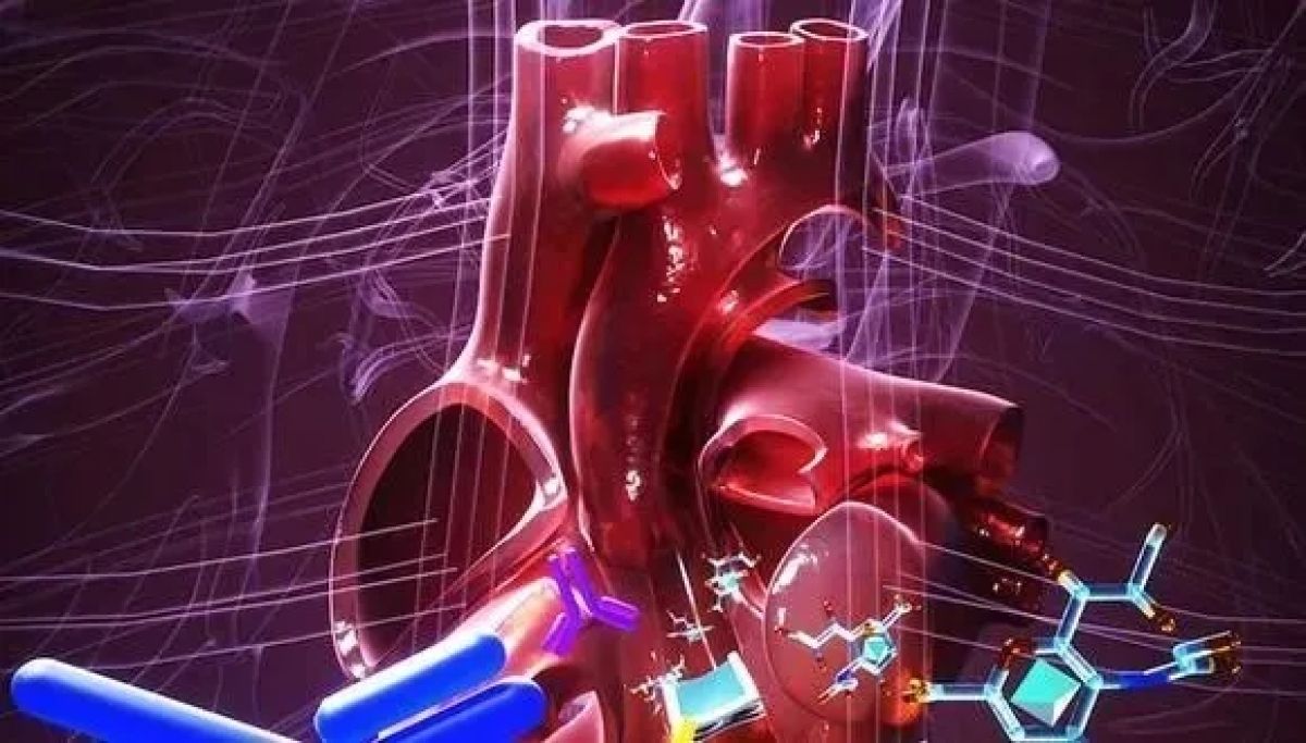
An international study led by a researcher from Tel Aviv University offers a novel technology that can assist many patients implanted with bioprosthetic heart valve by avoiding additional complicated replacement surgery. Today, many heart patients implanted with such valves are forced to replace it ten years later due to calcification of valve tissue. The researchers have now been able to show that by genetically engineering the biological component in the valve, immunological attack and calcification risk can be avoided – thereby offering next-generation durable bioprosthetic heart valves.

This technological development stems from the EU-funded TRANSLINK consortium consisting of 14 members from Europe, USA and Canada and led by Dr. Vered Padler-Karavani from The Shmunis School of Biomedicine and Cancer Research, The George S. Wise Faculty of Life Sciences at Tel Aviv University. In this study, the researchers investigated close to 1700 patients with ~5000 blood samples covering almost 15 years since implantation. The study was published in the prestigious journal Nature Medicine.

The researchers explain that patients can be implanted with either a mechanical heart valve that last long but requires daily administration of anticoagulants that can lead to life-threatening hemorrhages, or bioprosthetic heart valves (made from bovine, porcine or equine tissue) allowing patients to live a reasonably normal life, but those are commonly destroyed after a decade requiring replacement.

Getting Rid of Foreign Sugars

Dr. Padler-Karavani: “since bioprosthetic heart valves are made of animal tissues, we hypothesized they contain foreign non-human sugars (Neu5Gc and alpha-Gal) that are attacked by the human immune system, which then mediate the calcification that lead to structural valve deterioration. Indeed, in our research we proved that this was the reason and even suggested an implementable solution.

“We discovered that all bioprosthetic heart valve patients developed an immune response against the foreign sugars in the valves. We could clearly see an increase in antibody responses against these sugars in implanted patients, as early as one month after implantation, some lasting even two years later. We also found that some of the patients showed signs of calcification as early as two years post implantation”.
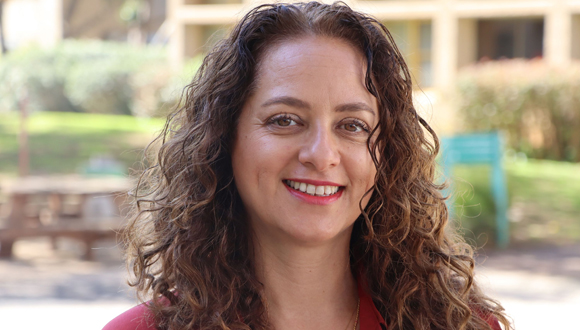
 

Dr. Vered Padler-Karavani

Anu Paul, a postdoc in the lab, also showed that the foreign sugars and the antibodies attacking them were found on calcified bioprosthetic heart valves explanted from patients some 10 years after implantation. Additionally, the dietary non-human sugar Neu5Gc and the antibodies against it were also found on explanted calcified native valves (the original malfunctioning human valves that needed to be replaced with a bioprosthesis). Since this sugar cannot be produced in the human body, it most likely accumulates on these valves from diet rich in red meat and dairy products where it is abundant. Therefore, it is possible that red meat diet mediates the initial need for valve replacement. The researchers also confirmed in a human-like animal model that antibodies against the foreign sugars indeed mediate calcification of tissues used for production of bioprosthetic heart valves.

Furthermore, the option to employ genetic engineering to resolve the problem was examined. For this purpose, the consortium created genetically modified porcine (pigs) that do not express the sugars foreign to humans. In Dr. Padler-Karavani’s lab the researchers found that in a human-like animal model that engineered tissue lacking the foreign sugars significantly had reduced calcification even in the presence of antibodies against the sugars and can therefore increase the durability of bioprosthetic heart valves made of such tissues.

“This study marks breakthrough technology in the field of bioprosthetic heart valves and provides deep understanding of the mechanisms leading to structural valve deterioration. These findings can lead to a dramatic improvement in the quality of life of many heart patients. Now, it would be interesting to study whether vegetarians or people who consume only small amounts of red meat and dairy have lower probability of heart valve calcification, and if this could perhaps be associated with low levels of antibodies against these foreign sugars. In the future, it may also be possible to devise a modified diet to reduce the risk or to actually produce biological valves from the tissues of engineered animals that do not contain the sugars at all”, Dr. Padler-Karavani summarizes.

Israeli Breakthrough in Treating PTSD

World first: TAU-led team shows success of oxygen therapy in alleviating symptoms of PTSD in military veterans.

Researchers from Tel Aviv University and Israel’s Shamir Medical Center were able to successfully relieve the symptoms of post traumatic stress disorder (PTSD) in military combat veterans using a new protocols of hyperbaric oxygen therapy (HBOT). In a controlled clinical trial involving Israeli army veterans who suffered from treatment-resistant PTSD, the approach demonstrated significant improvement in all classes of symptoms.

According to the World Health Organization (WHO), almost 4% of the global population, and 30% of all combat soldiers, develop PTSD. 

Hyperbaric medicine involves treatments in a pressurized chamber where atmospheric pressure is higher than sea-level pressure and the air is rich with oxygen. Considered a safe form of treatment, hyperbaric oxygen therapy is already used for a range of medical conditions. Evidence gathered in recent years indicates that special hyperbaric protocols can improve the supply of oxygen to the brain, thereby enhancing the generation of new blood vessels and neurons. It must be noted that HBOT treatments require the evaluation and supervision of qualified physicians. Moreover, for medical indications it should be given using a certified chamber with appropriate quality assurance using the exact studied treatment protocols. 

The breakthrough research was led by Prof. Shai Efrati, Dr. Keren Doenyas-Barak, and Dr. Amir Hadanny of Tel Aviv University’s Sackler Faculty of Medicine and Sagol School of Neuroscience in cooperation with Shamir Medical Center. The team also included Dr. Ilan Kutz, Dr. Merav Catalogna, Dr. Efrat Sasson, Gabriela Levi and Yarden Shechter of Shamir Medical Center.

Unloading Pain for a Better Future

The study included 35 combat veterans of the Israel Defense Forces (IDF) who suffered from PTSD that was resistant to both psychiatric medications and psychotherapy. 

“The veterans were divided into two groups: one group received hyperbaric oxygen therapy while the other served as a control group,” explains Dr. Keren Doenyas-Barak of Shamir Medical Center. “Following a protocol of 60 treatments improvement was demonstrated in all PTSD symptoms, including hyper-arousal, avoidance, and depression. Moreover, both functional and structural improvement was observed in the non-healing brain wounds that characterize PTSD. We believe that in most patients, improvements will be preserved for years after the completion of the treatment.”

“This study gives real hope to PTSD sufferers. For the first time in years the study’s participants, most of whom had suffered from severe PTSD, were able to leave the horrors behind and look forward to a better future.”

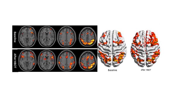
Illustration: Clinical example of functional brain imaging by fMRI.  The reduced brain activity in the frontal lobes of the brain (responsible among others for emotional regulation and executive functions) and in hippocampus (responsible for memories functions) is improved after Hyperbaric Oxygen Therapy (HBOT).

Emotional Trauma Can Cause Physical Damage

“Today we understand that treatment-resistant PTSD is caused by a biological wound in brain tissues, which obstructs attempts at psychological and psychiatric treatments,” explains TAU Prof. Shai Efrati. “With the new hyperbaric oxygen therapy protocols, we can activate mechanisms that repair the wounded brain tissue. The treatment induces reactivation and proliferation of stem cells, as well as generation of new blood vessels and increased brain activity, ultimately restoring the functionality of the wounded tissues. Our study paves the way to a better understanding of the connection between mind and body.”

“Our results indicate that exposure to severe emotional trauma can cause organic damage to the brain,” says Prof. Efrati. “We also demonstrate for the first time that direct biological treatment of brain tissues can serve as a tool for helping PTSD patients. Moreover, our findings may be most significant for diagnosis. To date, no effective diagnostic method has been developed and diagnosis of PTSD is still based on personal reports which are necessarily subjective – leading to many clashes between the suffering veterans and the authorities responsible for treating them. Think of a person who comes to the emergency room with chest pains. The pain might be caused by either a panic attack or a heart attack, and without objective EKG and blood tests, the doctors might miss a heart attack. At present we are conducting continuing research in order to identify the biological fingerprint of PTSD, which can ultimately enable the development of innovative objective diagnostic tools.”


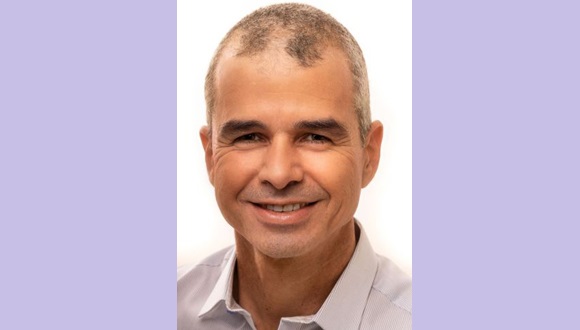
Prof. Shai Efrati

Prof. Efrati is an Associate Professor at TAU and director of the Sagol Center for Hyperbaric Medicine and Research at Shamir Medical Center. He is also the co-founder and Chair of the Medical Advisory Board at Aviv Scientific LTD, a company that applies the hyperbaric oxygen therapy protocols developed from his team’s research to enhance the brain and body performance of aging adults. 

Discovery May Enable Early Diagnosis of Alzheimer’s

Can your sleep predict your chance of developing the disease?

A new study at Tel Aviv University revealed abnormal brain activity that precedes the onset of Alzheimer’s first symptoms by many years: increased activity in the hippocampus, a region of the brain which plays a key role in memory processes, during anesthesia and sleep, resulting from failure in the mechanism that stabilizes the neural network. The researchers believe that the discovery of this abnormal activity during specific brain states may enable early diagnosis of Alzheimer’s, eventually leading to a more effective treatment of a disease that still lacks effective therapies.

The study was published in the prestigious scientific journal Cell Reports, and led by Prof. Inna Slutsky and doctoral students Daniel Zarhin and Refaela Atsmon from the Sackler Faculty of Medicine and the Sagol School of Neuroscience at Tel Aviv University.

According to Prof. Inna Slutsky, innovative imaging technologies developed in recent years have revealed that amyloid deposits, a hallmark of Alzheimer’s disease pathology, are formed in patients’ brains as early as 10-20 years before the onset of typical symptoms such as memory impairment and cognitive decline. Unfortunately, most efforts to treat Alzheimer’s disease have failed. She believes that if we could detect the disease at the pre-symptomatic stage, and keep it in a dormant phase for many years, this would be a tremendous achievement in the field. Identifying a signature of aberrant brain activity in the pre-symptomatic stage of Alzheimer’s and understanding the mechanisms underlying its development she says may be a key to effective treatment.

Additional participants in the study include: Dr. Antonella Ruggiero, Halit Baeloha, Shiri Shoob, Oded Scharf, Leore Heim, Nadav Buchbinder, Ortal Shinikamin, Dr. Ilana Shapira, Dr. Boaz Styr, and Dr. Gabriella Braun, all from Prof. Slutsky’s laboratory. Collaborations with the laboratory teams of Prof. Yaniv Ziv of the Weizmann Institute, and Prof. Yuval Nir of TAU were essential for the project. Prof. Tamar Geiger, Dr. Michal Harel, and Dr. Anton Sheinin of Tel Aviv University, as well as researchers from Japan, also contributed to the study.

The researchers used animal models for Alzheimer’s, focusing on the hippocampal region of the brain, which is known to be impaired in Alzheimer’s patients. At first, they measured cell activity in the hippocampus when the model animal was awake and active. For this, they used advanced methods that measure brain activity at a resolution of single neurons.

High Neuronal Activity – Also During Sleep

“It is known that neuronal activity of the hippocampus decreases during sleep in healthy animals,” explains Refaela Atsmon. However, when she examined model animals in early stages of Alzheimer’s, she found that their hippocampal activity remained high even during sleep. This is due to a failure in the physiological regulation, which she says has never before been observed in the context of Alzheimer’s disease.

Daniel Zarhin found similar dysregulation in model animals under anesthesia: neuronal activity did not decline, the neurons operated in a manner that was too synchronized, and a pathological electrical pattern was formed, similar to ‘quiet’ seizures in epileptic patients.

The researchers found that brain states that block responses to the environment – such as sleep and anesthesia – expose abnormal activity which remains hidden when the animal is awake, and this happens before the symptoms of Alzheimer’s disease are observed.

Prof. Slutsky’s explains that even though this abnormal activity can be detected during sleep, it is much more frequent under anesthesia. Therefore, she says, it would be important to test whether short anesthesia can be used for early diagnosis of Alzheimer’s disease.

Defective Stabilizing Mechanisms

The researchers proceeded to ask what causes the abnormality. To this end, they relied on findings from previous studies from Prof. Slutsky’s laboratory and other researchers on homeostasis of neural networks: each neural circuit has a set point of activity, maintained by numerous stabilizing mechanisms. These mechanisms are activated when the balance is disturbed, restoring neuronal activity to its original set point.  

Is a disruption of these mechanisms the main cause of deviant brain activity during sleep and anesthesia in Alzheimer’s disease animal models? To test this, Dr. Antonella Ruggiero examined the effect of various anesthetics on neurons grown on a chip. She found that they lower the set point of neuronal activity. While in healthy neural networks this activity remained low over time, in neural networks expressing genetic Alzheimer’s mutations, the lowered set point recovered quickly, despite the presence of anesthetics.

The researchers now sought to examine a potential drug for the impaired regulatory mechanism. According to Prof. Slutsky, the instability in neuronal activity found in the study is known from epilepsy. In a previous study Prof. Slutsky’s team discovered that an existing drug for multiple sclerosis may help epilepsy patients by activating a homeostatic mechanism that lowers the set point of neural activity. Doctoral student Shiri Shoob examined the effect of the drug on hippocampal activity in the animal model for Alzheimer’s and found that also in this case the drug stabilizes activity and reduces pathological activity observed during anesthesia.

Proceeding Towards Clinical Trials

“The results of our study may help early diagnosis of Alzheimer’s, and even provide a solution for instability of neuronal activity in Alzheimer’s disease,” says Prof. Slutsky. Firstly, we discovered that anesthesia and sleep states expose pathological brain activity in the early stages of Alzheimer’s disease, before the onset of cognitive decline. We also proposed the cause of the pathological activity – failure of a very basic homeostatic mechanism that stabilizes electrical activity in brain circuits. Lastly, we showed that a known medication for multiple sclerosis suppresses this type of anesthesia-induced aberrant brain activity,” she concludes.

The researchers now plan to collaborate with medical centers in Israel and worldwide to test whether the mechanisms discovered in animal models can also be identified in patients with early-stage Alzheimer’s disease. For this purpose, they propose to incorporate EEG monitoring into surgical procedures, to measure brain activity of patients under anesthesia. They hope that their findings will promote early diagnosis and drug development for the most common form of late-onset dementia.

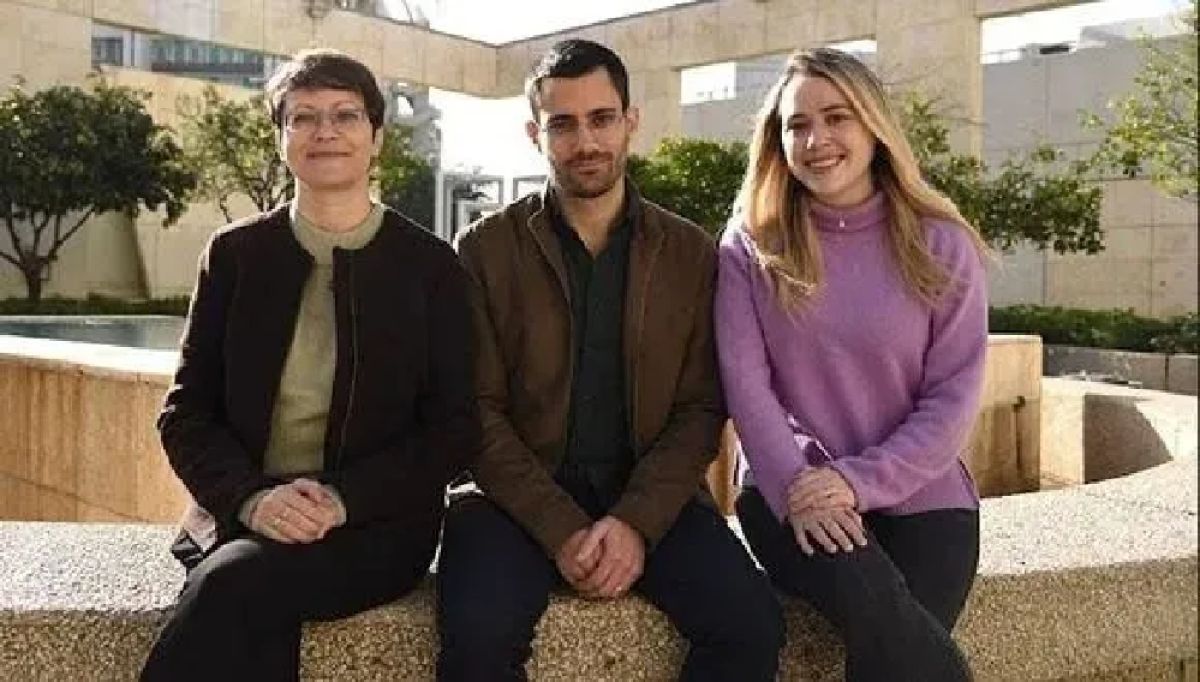
Featured image: The Research Team (from left to right): Prof. Inna Slutsky, Daniel Zarhin and Refaela Atsmon (Photo: Dr. Tal Laviv)

TAU Breakthrough Offers New Hope to Help People With Paralysis Walk Again

Researchers successfully engineer world’s first 3D human spinal cord tissue transplant.

Paralysis from spinal injury has long remained untreatable. Could scientific developments get people affected on their feet again sooner than imagined? In a worldwide first, Tel Aviv University researchers have engineered 3D human spinal cord tissues and implanted them in a lab model with long-term chronic paralysis, demonstrating high rates of success in restoring walking abilities. Now, the researchers are preparing for the next stage of the study, clinical trials in human patients. They hope that within a few years the engineered tissues will be implanted in paralyzed individuals enabling them to stand up and walk again.

How to Reverse Spinal Injury?

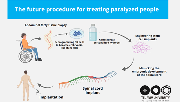
“Our technology is based on taking a small biopsy of belly fat tissue from the patient,” explains Prof. Tal Dvir who’s research team led the study. “This tissue, like all tissues in our body, consists of cells together with an extracellular matrix comprising substances like collagens and sugars. After separating the cells from the extracellular matrix we used genetic engineering to reprogram the cells, reverting them to a state that resembles embryonic stem cells – namely cells capable of becoming any type of cell in the body.”


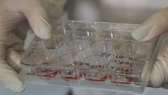
Petri dish with tissue samples (Photo: Sagol Center for Regenerative Biotechnology)

From the extracellular matrix the researchers produced a personalized hydrogel, that would evoke no immune response or rejection after implantation. They then encapsulated the stem cells in the hydrogel, and in a process that mimics the embryonic development of the spinal cord, turned the cells into 3D implants of neuronal networks containing motor neurons.


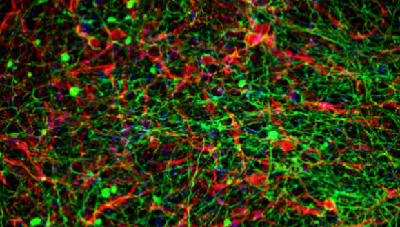
Neural net (Photo: Sagol Center for Regenerative Biotechnology)

The human spinal cord implants were then implanted in two different groups of lab models: those who had only recently been paralyzed (the acute model) and those who had been paralyzed for a long time (the chronic model) – equivalent to one year in human terms. Following the implantation, 100% of the lab models with acute paralysis and 80% of those with chronic paralysis regained their ability to walk.

The groundbreaking study was led by Prof. Tal Dvir’s research team at the Sagol Center for Regenerative Biotechnology, The Shmunis School of Biomedicine and Cancer Research, and the Department of Biomedical Engineering at Tel Aviv University. The team at Prof. Dvir’s lab includes PhD student Lior Wertheim, Dr. Reuven Edri, and Dr. Yona Goldshmit.  Other contributors included Prof. Irit Gat-Viks from the Shmunis School of Biomedicine and Cancer Research, Prof. Yaniv Assaf from the Sagol School of Neuroscience, and Dr. Angela Ruban from The Stanley Steyer School of Health Professions at the Sackler Faculty of Medicine, all at Tel Aviv University. The results of the study were published in the prestigious scientific journal Advanced Science.

Visualization of the next stage of the research – human spinal cord implants for treating paralysis (Photo: Sagol Center for Regenerative Biotechnology)

Getting Patients Suffering from Paralysis Back on Their Feet

Encouragingly, the model animals underwent a rapid rehabilitation process, at the end of which they could walk quite well. This is the first instance in the world in which implanted engineered human tissues have generated recovery in an animal model for long-term chronic paralysis – which is the most relevant model for paralysis treatments in humans.

“Our goal is to produce personalized spinal cord implants for every paralyzed person, enabling regeneration of the damaged tissue with no risk of rejection,” says Prof. Dvir.

Based on the revolutionary organ engineering technology developed at Prof. Dvir’s lab, he teamed up with industry partners to establish Matricelf (matricelf.com) in 2019. The company applies Prof. Dvir’s approach in the aims of making spinal cord implant treatments commercially available for persons suffering from paralysis.

Prof. Dvir, head of Sagol Center for Regenerative Biotechnology, concludes: “We hope to reach the stage of clinical trials in humans within the next few years, and ultimately get these patients back on their feet. The company’s preclinical program has already been discussed with the FDA. Since we are proposing an advanced technology in regenerative medicine, and since at present there is no alternative for paralyzed patients, we have good reason to expect relatively rapid approval of our technology.” 


The research team (from left to right): Dr. Yona Goldshmit, Prof. Tal Dvir and Lior Wertheim  (Photo: Sagol Center for Regenerative Biotechnology)


Ketogenic Diet Likely to Reduce Damage from Traumatic Brain Injuries

TAU-led study finds diet improves spatial and visual memory, lowers signs of brain inflammation, reduces neuronal death and slows down cellular aging.

Traumatic Brain Injury (TBI) is a leading cause of death and long-term disability in the developed world. It is estimated that every year over 10 million people worldwide suffer from traumatic brain injury as a result of head injuries caused by a hard object, a blow, an explosion, road accidents, sports injuries, etc. Such traumas can lead to physical, cognitive, behavioral and emotional damage and is also a risk factor for diseases such as Alzheimer’s and Parkinson’s.

At this point, despite the high frequency of brain injuries, there is no proven effective treatment that can help those suffering from this injury.

A new international study piloted by the Tel Aviv University determines that a ketogenic diet may reduce the effects of brain damage after traumatic injury. The study indicates that the diet improves spatial memory and visual memory, lowers brain inflammation indices, causes less neuronal death and slows down the rate of cellular aging.

The study was led by Prof. Chaim (Chagi) Pick, Director of the Sylvan Adams Sports Institute and a member of the Sagol School of Neuroscience, and Ph.D. student Meirav Har-Even Kerzhner, a registered dietitian and brain researcher, both from the Sackler Faculty of Medicine at Tel Aviv University. The findings were published in Scientific Reports, a syndicate journal from the publishers of Nature.

What does a ketogenic diet consist of?

A ketogenic diet which involves changes in the consumption of common foods is based on high-fat percentages and aims to mimic a state of fasting. As part of the diet, the intake of foods that contain carbohydrates (e.g., bread, sugar, grains, legumes, snacks, pastries and even fruits) are significantly restricted and, at the expense of this restriction, high-fat products such as meat, fish, eggs, avocado, butter etc. – are eaten.

This is a diet that can be continued for extended periods of time. The diet causes an increased production of ketone bodies in the liver that are used to generate energy. These ketone bodies are transferred via the bloodstream to the brain providing optimal nourishment.

The diet has been used as a treatment in Israel and around the world for almost 100 years, among children with epilepsy, while in recent years, the ketogenic diet has become popular among those who want to lose weight. It is important to note that, due to the significant nutritional restrictions, it is necessary to consult with a professional such as a doctor or a registered dietitian.

Inspiring Hope

In the study, conducted on model animals, the researchers identified that the ketogenic diet greatly improves the patient’s brain function. For this purpose, the researchers used advanced methods that included, among other things, behavioral-cognitive tests, biochemical tests and immunohistochemical cell staining (a technique in biology for the detection and placement of proteins in a cross section of tissue). The mechanism by which a ketogenic diet succeeds in benefiting the results of brain damage has not yet been fully revealed, but studies show that it has an antioxidant and metabolic effect on mitochondria (essential organelles in the cell, the its primary function of which is energy production and respiration), lowers free radical production and raises ATP (a major molecule in cellular biochemical channels). 

“The findings were unequivocal and showed that the ketogenic diet improves spatial memory and visual memory, lowers indices of inflammation in the brain and in addition, also slows the rate of cellular aging,” says Prof. Pick. “These results may open the door to further research that will inspire hope for those suffering from traumatic brain injuries, and their family members.”

Featured image: Prof. Chaim Pick and Ph.D. student Meirav Har-Even Kerzhne

Viruses and Game Theory

TAU researchers discover new mechanism for communication between viruses and bacteria.

Phages are viruses that attack bacteria. Many phages can exist in one of two states: active (lysis), in which the phages attack and destroy bacteria, or dormant, in which they remain passive within the bacteria, replicating themselves but doing no damage (lysogeny). Phages of this type must decide whether to be active or dormant every time they infect a new host. If they decide to be dormant for a time, they must also decide when to ‘wake up’ and attack. As in all dilemmas, it’s important to base the decision upon solid, reliable information.

Researchers at Tel Aviv University have discovered that just like humans with Game Theory, phages weigh all their options and make an informed decision on whether it is time to exit the dormant state and attack their bacterial host. The study was led by Prof. Avigdor Eldar of The Shmunis School of Biomedicine and Cancer Research at Tel Aviv University, together with his students and partners from the Weizmann Institute of Science. The paper was published in December 2021 in the journal Nature Microbiology.

According to the researchers, it has been assumed for some time that a phage bases its decision to exit the dormant state on information regarding the condition of its bacterial host: when the host shows signs of substantial DNA damage (death throes, so to speak), it is in the phage’s interest to leave it and try to infect other bacteria.

The new study discovered an additional mechanism of communication between bacteria and phages: apparently, some phage families have developed a more complex decision-making strategy, a kind of ‘phage game theory’, in which the phage receives information not only from its own host but also from neighboring bacteria.

What’s Going On in The Neighborhood?

Prof. Eldar explains: “When a phage is dormant within a bacterial cell, it forces its host to constantly produce small communication molecules called arbitrium, to which the phage listens via a special receptor. Thus, the presence of high levels of these molecules indicates that neighboring bacteria also contain phages. When this happens, even if its own host exhibits DNA damage, the phage refrains from becoming active. Since every bacterium can only host one dormant phage, the phage makes an informed decision: it’s better to let the host try to repair itself than to ‘betray’ it, since all neighboring bacteria are already taken.”

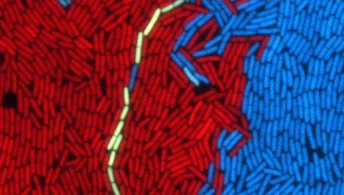
Prof. Eldar and his team used a range of genetic and biomolecular methods to track the biochemical communication signals passing between the bacteria and phages. In a former study they used a fluorescent marker to show that communication methods used by phages, as well as a large family of similar communication systems (know generally as ‘quorum sensing’) are used only to get signals from close neighbors. “Essentially, the bacteria have developed two separate communication systems – one for long-range communication, and the other for short distances only, used to sense the state of their immediate neighbors,” says Prof. Eldar. “In the phage’s case, it controls communication, and is only interested to know whether its close neighbors, which it might easily infect, are already occupied.”

Prof. Eldar concludes: “Several years ago, Prof. Rotem Sorek and his team at the Weizmann Institute identified communication between phages for the first time. Such systems had been known to exist between other molecular parasites hosted by bacteria (called plasmids). Our new discovery is the fact that phages use communication even in their dormant state. We have identified components critical for understanding how phages combine information about their host’s condition with information about their neighbors. This is one more important step on the way to deciphering the communication and ‘behavioral economics’ of viruses. Phages have an excellent ability to process information and make the right decision to ensure optimal survival. It will be interesting to see whether viruses residing in more complex organisms but facing similar decisions have also developed comparable systems of communication.”

What’s The Link Between Electrical Voltage and Brain Adaptability?

New study finds direct and significant link between changes in G-protein-coupled receptors and the brain’s ability to adapt to external changes.

Our brain has a large amount of G protein-coupled receptors (GPCR). Activation of these proteins causes a chain of chemical reactions within the cell. These proteins are very common in the brain and are involved in almost every brain activity, such as learning and memory. The nerve cells in which GPCRs are common, experience changes in their electrical voltage.

20 years ago, it was unexpectedly discovered that GPCRs are voltage-dependent, meaning that they sense the changes in the electrical voltage of nerve cells and change their function. However, to date, it has not been clarified whether the voltage dependence of GPCR proteins has a physiological significance that affects brain activity, our perception, and behavior. In fact, the scientific mindset was that this voltage dependence has no physiological significance.

The study, published recently in the prestigious journal Nature Communications, was conducted by Dr. Moshe Parnas and his team from the Sackler Faculty of Medicine and the Sagol School of Neuroscience at Tel Aviv University.

The Protein that Influences our Sense of Smell

Dr. Parnas and his team investigated, by means of the olfactory system of the fruit fly, whether the voltage dependence of GPCRs is important for brain function. To this end, the researchers decided to focus on one receptor from the G protein-coupled receptor family called “Muscarinic Type A”. This protein is involved, among other things, in habituation to an odor, a process in which the intensity of the reaction to the odor decreases as a result of continuous exposure to it. Thanks to this mechanism, a few minutes after entering a room containing a distinct odor – we stop smelling it.

Dr. Parnas explains: “Nerve cells are able to communicate with each other and brain flexibility is expressed in the ability of nerve cells to set up new connections with each other and change existing connections – and thus influence behavior. Muscarinic Type A protein is involved in strengthening the bond between nerve cells, and strengthening of this bond causes fruit flies to get used to the odor and indicates normal brain flexibility.”

During the course of the study, the researchers were able to neutralize the voltage sensor of the “Type A” Muscarinic protein by means of genetic editing, and thus eliminate its dependence on the electrical voltage of the nerve cell. The researchers found, by applying molecular, genetic and physiological methods, that disabling the voltage sensor actually causes uncontrolled brain flexibility and consequently the process of excessive and uncontrolled habituating to an odor.


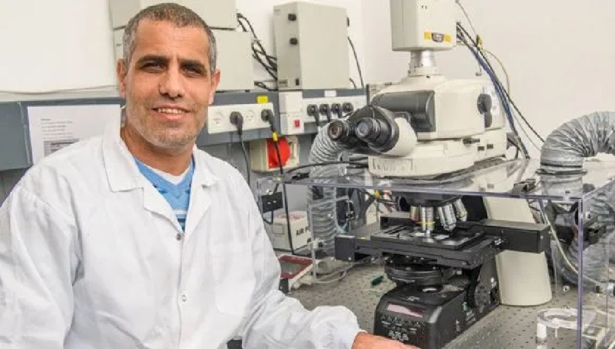
Dr. Moshe Parnas

Control Mechanism Uncovered

Dr. Parnas adds: “We found that the receptor in question is very much involved in strengthening the intercellular bond in the brain, much more than what we thought. When we turned off its voltage sensor, the connection between the nerve cells became too strong.”

According to Dr. Parnas, “These findings change our perception of G-protein-coupled receptors. To date, no reference has been made to the effect of electrical voltage on their function and its implications on brain flexibility and conduct. These receptors are involved in many systems and brain diseases and we have now discovered a control mechanism upon which an attempt at drug treatment can be based.”

“Following this, we are continuing to investigate additional receptors. It is reasonable to assume that their dependence on the electrical voltage is important in other systems and not only in the olfactory system [i.e. the bodily structures that serve the sense of smell].”

This study by Dr. Parnas is a follow-up to a study conducted by his parents about two decades ago, which focused solely on the protein level. The current study by Dr. Parnas and his team advances to the next stage, connecting molecules, brain and conduct and indicating, for the first time, that eliminating their ability to sense electrical voltage affects brain activity and our ability to optimally adapt to the environment.

As Pandemic Persists, TAU Forges Ahead with COVID-19 Research on All Fronts

The University’s Center for Combating Pandemics leads research efforts in a range of fields.

As the COVID-19 pandemic nears the end of a second year, Tel Aviv University is maintaining its fast pace of scientific discovery in the global battle against coronavirus.  

Building on the University’s innovation record, TAU in 2020 launched the Center for Combating Pandemics. The Center harnesses the collective power of academia, hospitals, government and industry to promote research and guide government policy. It is the first entity of its kind in Israel, and potentially the world.  

Members of the Center include more than 100 research groups comprised of top TAU experts from all scientific disciplines across campus. Among them, over 60 researchers have, until now, conducted projects related to COVID-19. 

Academic Powerhouse

To date, the Center’s members have published findings from dozens of COVID-19 studies. Select examples include: 

Prof. Noam Shomron (Medicine) spearheaded a novel approach to remote monitoring of COVID-19 patients that flags early cardiovascular warnings of worsening health. More here.  

Prof. Shoshana Shiloh (Social Sciences) and team devised a model to determine the psychological and social factors that predict who will voluntarily take the COVID-19 vaccine.  

Prof. Dan Yamin’s (Engineering) lab developed a “big data” COVID-19 detection technology that could be used to reduce viral spread and help shape more efficient testing policy. More here.  

Prof. Isaac Sasson (Engineering) led a study to help improve understanding of COVID-19 mortality and age in countries with limited data. More here.  

A team led by Prof. Saharon Rosset (Exact Sciences) built a statistical model to better understand SARS-CoV-2 evolution and to predict future mutations of the virus, particularly those which lead to new variants. 

Prof. Aeyal Gross (Law) and team examined legal and public health implications of Israel’s “Green Pass” proof-of-vaccination rules under the country’s pandemic response. More here.  

Through big data methods, Prof. Tal Pupko’s (Life Sciences, Shmunis School) found that the swift implementation of lockdowns, rather than their strictness, was more effective in reducing COVID-19 mortality rates. More here.  

New Findings on the Horizon

Currently, the TAU researchers from the Center have approximately 30 ongoing coronavirus projects, including:  

Prof. Dan Peer (Life Sciences), TAU Vice President for Research & Development, is exploring best practices to advance the bourgeoning technology behind non-viral mRNA vaccines. 

Prof. Adi Stern’s (Life Sciences, Shmunis) lab has been studying the evolution of SARS-CoV-2, including the origin and behavior of the Omicron variant. More here.  

Prof. Ron Shamir (Exact Sciences) has teamed up with local hospitals to explore early predictors of deterioration in COVID-19 patients using machine-learning and “big data” methods. 

Prof. Yael Benyamini (Social Sciences) is leading a broad study on pregnancy and childbirth that surveyed the concerns of pregnant women about childbearing amid COVID-19. 

Prof. Miri Yemini (Humanities) & Dr. Efrat Blumenfeld (Arts) are leading an urban-sociological examination of remote teaching and learning during times of social distancing. 

Upcoming Events

In March, the Center plans to hold its first international conference to highlight recent COVID-19 research. The event will also recognize trailblazing research fellows and grant recipients. For registration and updates, visit: https://en-pandemics.tau.ac.il/save-the-date-first-tccp-convention .

Featured News: More than 100 research groups from all scientific disciplines across TAU campus are on the frontlines of the pandemic battle. (Photo: Moshe Bedarshi)

Medicinal Cannabis Oil Effective for Treating Autism

Treatment improves both behavioral and biochemical parameters of the disease.

Autism is a neurodevelopmental disease, and its main symptoms are social deficiencies and compulsive behaviors. Cases range from mild to severe, and causes are both genetic and environmental. Researchers at Tel Aviv University have successfully treated autism in animal models with medical cannabis oil, improving behavioral and biochemical parameters.

Advancing in Reverse Order

In about 1% of all autism cases, a mutation in a single gene, called Shank3, is associated. While testing of medicinal cannabis traditionally begins with humans, in the current study PhD student Shani Poleg and Prof. Daniel Offen of TAU’s Sackler Faculty of Medicine, Felsenstein Medical Research Center and Sagol School of Neuroscience used animal models with a mutation in Shank3 to test the effectiveness of cannabis oil for alleviating symptoms of autism. The results of the surprising study were published in Translational Psychology published by Nature.

“We saw that cannabis oil has a favorable effect on compulsive and anxious behaviors in model animals,” says Shani Poleg. “According to the prevailing theory, autism involves over arousal of the brain which causes compulsive behavior. In the lab, in addition to the behavioral results, we saw a significant decrease in the concentration of the arousing neurotransmitter glutamate in the spinal fluid – which can explain the reduction in behavioral symptoms.”

A Little Euphoria Makes All the Difference

Attempting to determine which components of cannabis oil alleviate symptoms of autism, the researchers found that THC, the main psychoactive compound in cannabis which is responsible for the euphoric sensation associated with its use, is effective in treating autism, possibly even in small quantities.

“Clinical trials testing cannabis treatments for autism usually involve strains containing very large amounts of CBD – due to this substance’s anti-inflammatory properties, and because it does not produce a sense of euphoria,” explains Poleg. “Moreover, the strains used for treating autism usually contain very little THC, due to apprehension regarding both the euphoria and possible long-term effects.”

“In the second stage of our study we inquired which active substance in cannabis causes the behavioral improvement, and were surprised to discover that treatment with cannabis oil that contains THC but does not contain CBD produces equal or even better effects – both behavioral and biochemical. Moreover, our results suggest that CBD alone has no impact on the behavior of model animals.”

Addressing Existing Misinformation

Prof. Daniel Offen says, “Since cannabis is not defined as a medication, trials have already been conducted in children and adolescents with autism – without any preliminary studies addressing issues like the effect of cannabis on biochemical processes in the brain, spinal fluid or blood, and who can benefit from which type of cannabis oil. There is a great deal of misinformation on the subject of medicinal cannabis and autism, and Shani Peleg’s doctoral project represents pioneering basic research with regard to treating autism with cannabis oil.”

“This is an initial study,” concludes Poleg. “But we hope that through our basic research we will be able to improve clinical treatments. Our study shows that when treating autism with medicinal cannabis oil there is no need for high contents of either CBD or THC. We observed significant improvement in behavioral tests following treatments with cannabis oil containing small amounts of THC and observed no long-term effects in cognitive or emotional tests conducted a month and a half after the treatment began.”

TAU Students Racing Towards a Greener Campus

As part of Tel Aviv University’s initiative to reduce its environmental footprint to help combat the global climate crisis, the Entrepreneurship Center rallied 73 students to the cause and held a hackathon aimed at generating innovative solutions for a greener and more sustainable campus. The event was organized in cooperation with Shlomo Meltzer Institute for Smart Transportation and TAU’s Student Union.

Students were challenged to come up with practical solutions to one out of two central issues for cutting down pollution and waste: reducing the daily car traffic to campus and encouraging the use of reusable dishware on campus.

Fifteen experts mentored the students throughout the competition, which also featured professional enrichment and industry insider lectures on how organizations are addressing environmental challenges today.

Vehicular Pollution Challenge Winners

First Place: TAUapt

The winning team worked under the assumption that most TAU students residing in Tel Aviv rely on public transportation and therefore, do not use a car. What could encourage more of this behavior?

By enabling more students to live in Tel Aviv, the number of vehicles commuting to campus could further decrease. The team conceptualized a house hunting app, similar in nature to the existing job hunting platform AllJobs – but for apartments – which sends its subscribers instant messages through WhatsApp about apartments that are vacating. Team members: Abedallah Barghouti and Ubaydah Wattad.    


The TAUapt Team receives the prize for winning the competition’s Vehicular Pollution Challenge

Second Place: BIVPIsrael

The team suggested that the main difficulty for drivers of electric bicycles and scooters is the need to carry around a helmet and battery throughout the day. The team proposed a solution for storage compartments in which both helmet and battery can be left, and the latter can be charged, using solar-generated electricity, while the vehicle remains locked nearby.

Third Place: The Hitchhikers

The team proposed a way to encourage carpooling to campus by offering benefits to students and faculty members. They decided to appeal to people’s pockets – not their ideology.

Reusable Dishware Challenge Winners

First Place: Tengo

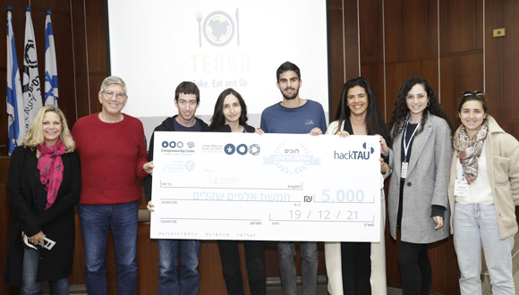
To cut down waste from single-use plastics, the Tengo team proposed a collaborative circular model based on the use of reusable dishware marked with a barcode. The reusable dishware is returned to collection boxes after use, where the users are credited. The dishware then gets   transported, cleaned and distributed back to campus restaurants. Team members: Chen Agassi, Idan Stark, Roi Farjun and Tali Aknin.

The Tengo Team receives the prize for winning the competition’s Reusable Dishware Challenge

Second Place: Go Clean Go Green

The team proposed a platform to incentivize the use of reusable dishware by awarding store credits for popular retailers. Through barcode-scanning technology on reusable cups and more, one earns redeemable points with every swipe.  

Third Place: TAUBIS

A game app that ranks businesses according to their degree of eco-friendliness. Students enjoy services similar to food delivery platforms like UberEats, Wolt or 10bis, accumulation of financial and ecological points.

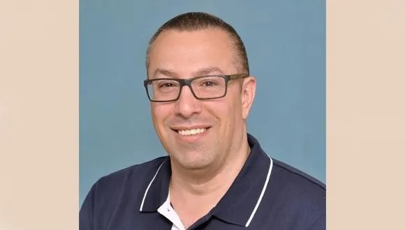
Yair Sakov, Head of TAU’s Entrepreneurship Center: “Sustainability and the circular economy are key issues promoted by the Entrepreneurship Center; there is no field more important than this to initiate and flood innovation.”

The Competition’s Judges

The judges in the competition included Knesset Member Prof. Alon Tal, former Head of Tel Aviv University’s Department of Public Policy; Miki Haimovich, Chairperson of the Heschel Center for Sustainability; Asi Schmelzer, Chairman of the Shlomo Group; Yuval Shani, VP of Technology and Innovation at Shlomo Group; Lior Hazan, Chairperson of TAU’s Student Union; Prof. Colin Price, Head of TAU’s Department of Environmental Studies; Prof. Hadas Maman, Head of the Environmental Engineering Program at TAU’s Faculty of Engineering; Dr. Ilit Oppenheim, Director at Shlomo Schmelzer Institute for Smart Transportation; Orlie Gruper, General Partner in Mobilitech Capital; Shani Raved, Global Operations Strategy Lead and Product Manager at Lime.  

Victoria

Tok Corporate Centre, Level 1,
459 Toorak Road, Toorak VIC 3142
Phone: +61 3 9296 2065
Email: [email protected]

New South Wales

Level 22, Westfield Tower 2, 101 Grafton Street, Bondi Junction NSW 2022
Phone: +61 418 465 556
Email: [email protected]

Western Australia

P O Box 36, Claremont,
WA  6010
Phone: :+61 411 223 550
Email: [email protected]