Israel’s reputation as “Startup Nation” is well-deserved, but its tech industry suffers from a lack of gender diversity. Hoping to bring in a wealth of untapped potential, Tel Aviv University’s Entrepreneurship Center created a semester-long hands-on workshop for women to help bring their startup ideas to fruition with the help of experienced mentors. One such project is Symbiosis CM, founded by student-and-mentor team Dr. Shira Burg and Varda Badet, which is creating a medical device that personalizes treatment of heart disease. The startup has raised $1.9M as of last year and won a number of competitions and grants including the Deep Tech Track of the Coller Startup Competition and a grant from the Israeli Innovation Authority.
The Problem
Originally trained as a veterinarian, Shira Burg worked for seven years alongside a small animal cardiologist in Israel, doing catheter procedures on dogs with congenital heart valve disease. There, she learned that one of the most common heart diseases, mitral valve disease, is difficult to treat because valve replacements are not fitted to individual heart sizes and replacement surgeries are invasive and costly. The disease occurs in aged dogs—and aged humans—in almost exactly the same way.
During this period, she began working with a surgeon who was attempting to create a mitral valve prosthetic, but discovered just how much heart anatomies differ from person to person.
“You can’t take something rigid and standard and expect it to fit every heart,” she says. “I realized that though we have personalized medicine in many fields such as drugs, diet, and genetics, we don’t have much personalization for medical devices. I saw an opening there for an improved mitral valve solution.”
Shira Burg: “I realized that though we have personalized medicine in many fields such as drugs, diet, and genetics, we don’t have much personalization for medical devices. I saw an opening there.”
Burg decided to pursue a PhD in cardiac electrophysiology at TAU’s Faculty of Medical and Health Sciences at the lab of Prof. Bernard Attali. There, she continued to think about her idea for a better mitral valve device. In 2020, she heard about a new workshop, Yazamiyot, for female graduate students at TAU’s Entrepreneurship Center. “I realized that if I could get accepted, I might be able to do something with my idea.” After a rigorous application process involving three separate interviews, Burg was one of 30 women admitted to the first course.
The Course
Yazamiyot, meaning “female entrepreneurs”, is an accelerator program targeting women master’s and PhD students. Participants work in small groups over one semester to establish a startup initiative in a supportive and empowering environment and are mentored through the process by successful women with industry experience.
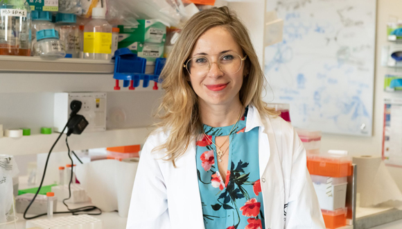
Burg’s mentor, TAU alumna Varda Badet, would go on to become her business partner. “With Varda, something just stuck. We had a connection,” Burg says.
Badet came from the finance sector, having served as the EVP at Bank Leumi for Risk Management during her many years in the field. Hoping to share her knowledge and connections, she took some courses to become a mentor at the Entrepreneurship Center. “There I found Shira, a very special entrepreneur. Every unsolved problem, she investigates and explores until it is solved. She is very motivated and ambitious, and I could see she was someone I wanted to work with.”
Yair Sakov: “At Tel Aviv University, we feel it is our responsibility to help drive change within society. Our mission at the Center is to empower students to come up with solutions to global challenges.”
During the semester, participants were brought face to face with the market realities that would determine whether their ideas were commercially feasible. They were taught how to inform themselves on target demographics and competitors, present their ideas to experts and the public, build a team, and more. They also heard from industry experts, from whom they could ask advice on their individual projects, and successful female entrepreneurs.
Yair Sakov, founder and managing director of the TAU Entrepreneurship Center, says: “At Tel Aviv University, we feel it is our responsibility to help drive change within our society. Our mission at the Center is to empower students to come up with solutions to global challenges by helping them develop an entrepreneurial mindset and by giving them tools and resources. In particular, we aim to uplift underrepresented communities, including women, who are still marginalized in the startup ecosystem. The Yazamiyot program was created to address that gender gap, and nearly every cohort since its inception has contributed to the establishment of new woman-led startups.”
Burg credits the workshop with giving her the tools to make her idea for a personalized heart valve device a reality, but says the most important thing she gained was her connection with Badet. Aside from teaching necessary business finance skills, Badet brought in resources in the form of her many industry connections and financial backing.
“As a mentor, you must believe in your entrepreneur completely and be ready to stick out the tough process with them. This product is worth that perseverance because it could save so many lives,” says Badet.
The Company
There is indeed great life-saving potential in addressing the lack of effective mitral valve disease solutions: in the US alone about 70% of the 4 million patients with the condition are unable to get proper treatment. Additionally, hospitals pay about $40 billion a year in treatment costs, with over 90% rehospitalization rates due to heart failure. Many companies have tried to address the problem, but their products are not widely approved and do not fit more than 20% of heart anatomies.
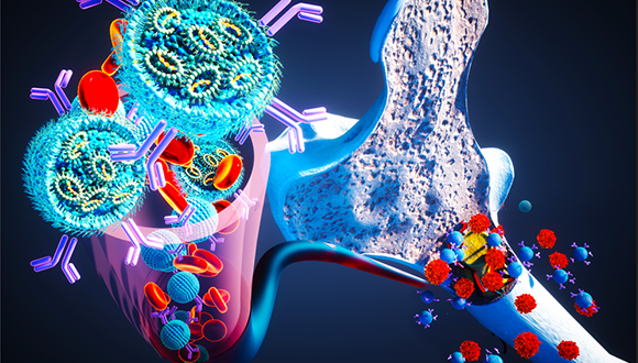
In a novel move, Symbiosis CM is developing a docking system that allows for real-time valve adjustment per an individual’s heart structure and minimally invasive procedures to treat the disease. The system is compatible with valves already on the market, making it accessible and opening the door to collaboration with other companies. The startup team already has a working prototype in preclinical trials on lab models.

A sample image of the heart valve docking system created by Symbiosis CM
Burg happily notes that her original plan to develop a solution for dogs, ideated during her time as a veterinarian, may one day also become reality: the product is small enough to work on animals as well as people.
The management side of things is also on track for success: Burg and Badet recently brought on a new CEO, Amir Weisberg, who has behind him 35 years of entrepreneurial experience, three exits in the medical field and an IPO on NASDAQ. “He was actually retired when we approached him, but after we presented to him, he decided to come out of retirement and sign on full-time to the company,” says Burg.
The Need for Women in Industry
Both Burg and Badet note that the Yazamiyot course is essential because of the difficulties of succeeding in the startup industry as a woman. Says Burg, “It’s a male-dominated world. We need to say it out loud. Especially in the spaces where I work, in the medical field and cardiology, it’s mostly men. When I came to specialists and investors, they would see a young woman and decide before I started speaking that I wasn’t to be taken seriously. That is why it is so important to push more women into entrepreneurship in general: so that people no longer question what we’re doing there.”
Adds Badet, “it makes me so glad to see and to help more women break into entrepreneurship.”
Shira Burg: “In Israel, female entrepreneurship is an unpolished diamond.”
“Israel has many undiscovered talents,” says Burg. “Female entrepreneurship, especially, is an unpolished diamond. If we expand programs such as Yazamiyot, amazing things will come from it.”
About the Entrepreneurship Center
Four the last four years, Tel Aviv University’s Entrepreneurship Center has provided students from across campus with the knowledge, tools, strategies and opportunities to create business and social ventures. It connects them with alumni, industry, government agencies and NGOs to generate and develop the next world-changing ideas. Over the Center’s first years of operations, it has achieved the following:
· 100 entrepreneurship courses, events and programs
· 12,000 student participants
· 92 startups
·$155 million in VC capital
·190 top industry mentors, most of them TAU alumni
By gradually expanding activities, the Center expects its to reach 4,500 students per year by 2029.