New TAU Treatment for Spinal Cord Injuries Shows Dramatic Recovery Results
Researchers developed an innovative therapy that reduces nerve cell damage after spinal cord injury and restored up to 80% of motor function in animal models
A new study led by Tel Aviv University offers real hope to millions worldwide affected by spinal cord injury (SCI), a devastating condition in which damage continues to spread after the initial trauma, often resulting in long-term and irreversible disability.
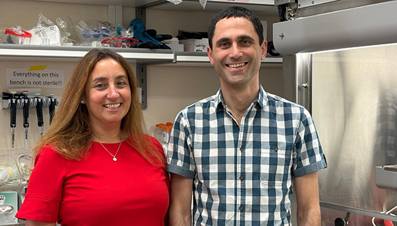
The study, recently published in the scientific journal Inflammation and Regeneration, presents an innovative therapeutic approach that reduces post-injury damage to nerve cells, reduces inflammation and scar formation, and therefore significantly improves functional recovery. The study was led by Dr. Angela Ruban from TAU’s Stanley Steyer School of Health Professions at the Gray Faculty of Medical & Health Sciences and the Sagol School of Neuroscience, together with Dr. Yona Goldshmit and students Josef Levin, Rosemary Lavender, Alexander Yakovchuk, Evgeny Banyas, and Ruth Baltovska. The findings were independently validated by a CRO as part of NeuroHagana’s preclinical development program, led by Dr. Amit Benbenishty.
Stopping the Damage Before It Spreads
The researchers explain that one of the main problems in SCI is a process occurring within minutes of the injury: the accumulation of a neurochemical called glutamate that further damages nerve cells, generating a local inflammatory response, degeneration that leads to scarring and extensive progressive damage. To date, no treatment has been approved by FDA/EMA to stop this process and prevent a permanent disability. This is where the new method comes in, introducing a novel therapeutic approach: Instead of attempting to block harmful activity in the brain, the researchers found a way to remove excess glutamate through the bloodstream in the first hours after injury.
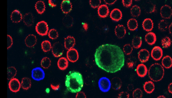
In experiments using animal models, the new treatment dramatically reduced post-injury glutamate levels, minimizing inflammation and nerve cell death, and preserving the structure of neural tissue, such as axons and synapses. Perhaps most impressive was the functional outcome: the treated animals showed marked improvement in walking and movement abilities within two days, achieving up to 80% of normal motor functioning two months after treatment – compared to around 30% in the untreated group.

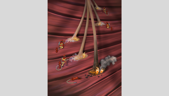
The spinal cord treatment process in mice
A Treatment Designed for Real Emergencies
Another important advantage is the wide therapeutic time window: the researchers found that the treatment remains effective when given as long as eight hours after injury – a timeframe considered realistic in real-world medical emergencies. Administered via a simple intravenous injection, the treatment may feasibly be provided by first responders in the field, thereby halting the damage cascade in its earliest stages.
Dr. Ruban notes that the importance of the study “is not only the functional improvement, but the very ability to impact secondary damage — for which no effective treatments have been discovered so far. This suggests a potential for halting the ‘chain reaction’ that aggravates patients’ condition, thereby preserving neural functions that would otherwise be lost. If we are able to confirm our results in humans, the new approach will represent a true paradigmatic shift – from supportive care alone to treatment that actually reduces and maybe even completely prevents the extent of the damage.”
Beyond Spinal Cord Injuries
Dr. Goldshmit, an expert in SCI treatment and rehabilitation, adds that this novel method can revolutionize treatment not only for SCI, but also for other brain injuries, caused for example by stroke or trauma. Significantly reducing neural damage, the new treatment can enable much more successful rehabilitation later on.
According to Dr. Ruban, the events of October 7 and the ensuing war have created an additional target for the study: head injuries resulting from blast waves. Equipped with encouraging preliminary results in head injury models, the researchers will now test the treatment for blast-induced head injury in lab models through collaboration with the Neurotechnology Department of Israel’s Ministry of Defense and TAU Professor Chaim Pick of the Sylvan Adams Sports Institute, Sagol School of Neuroscience, and Gray Faculty of Medical and Health Sciences.
Meanwhile, Ramot, TAU’s Technology Transfer Company, has established a commercial company to implement the breakthrough technology that redefines the treatment of both SCI and traumatic brain injuries (TBI): a simple intravenous injection with a wide therapeutic time window that reduces disability, improves quality of life, and significantly lowers costs for healthcare systems.
Dr. Ruban concludes: “Our main findings show that it is possible to intervene in the harmful process occurring immediately after injury — not just try to deal with its consequences after the fact. By reducing excess glutamate, we were able to protect nerve cells and significantly improve motor/cognitive functions in multiple preclinical models. If we can obtain similar results in humans, this study can potentially revolutionize the therapeutic approach to SCI and other neurological conditions. Together with other advanced medical and rehabilitation technologies being introduced into clinics, our innovation can help create a future in which SCI no longer condemns individuals to life in a wheelchair.”