Tag: Medicine

First 3D-bioprinting of entire active tumor

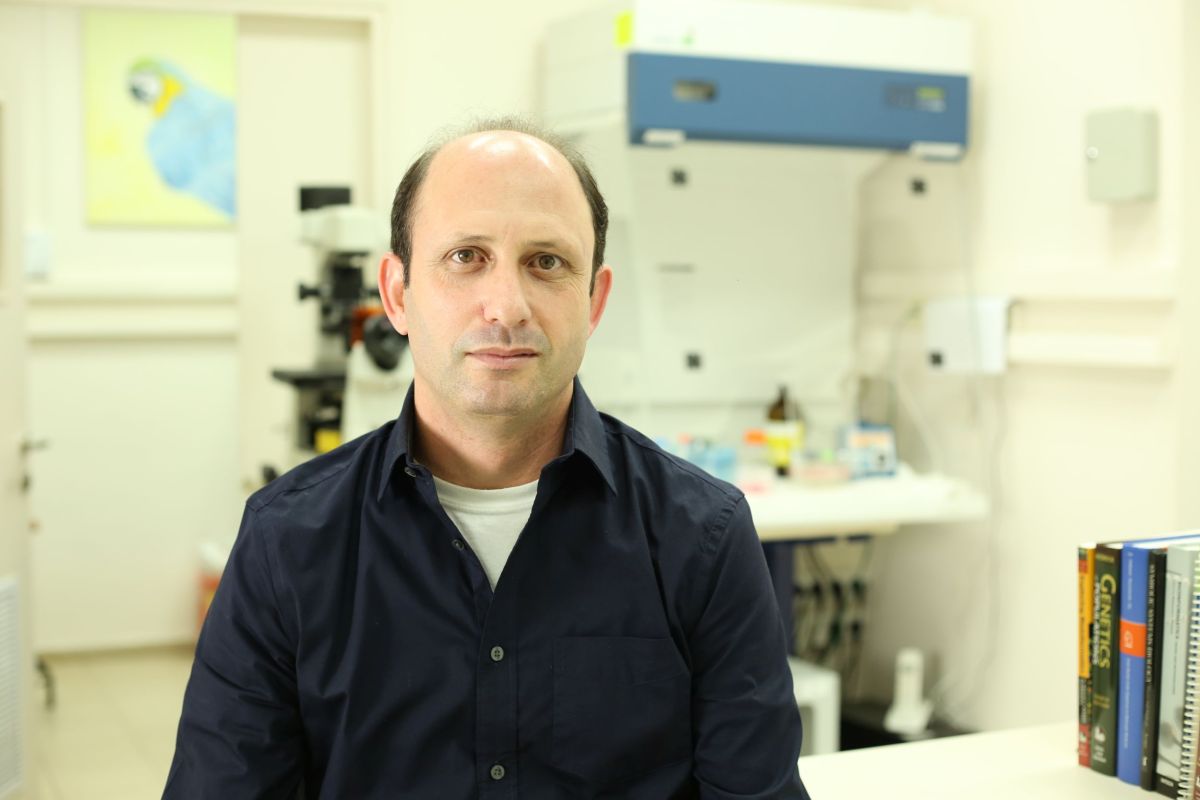
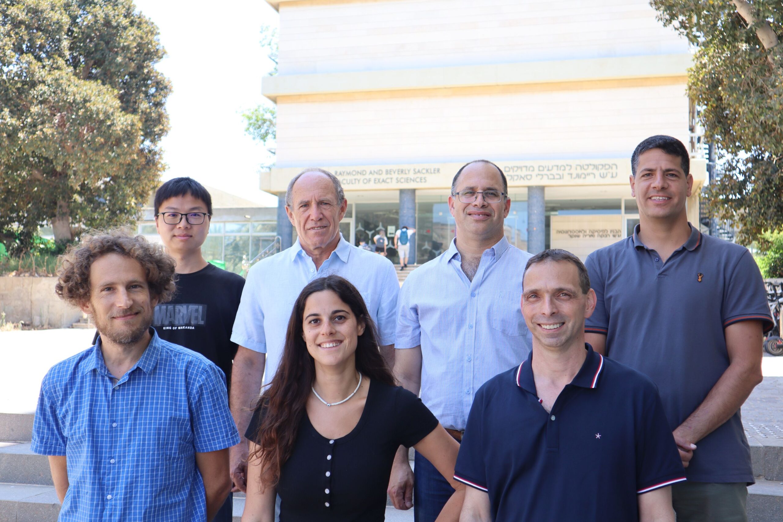
Research team(Left to right): Eilam Yeini, Prof. Satchi-Fainaro and Lena Neufeld

Scientific breakthrough in the battle against cancer

The 3D print of glioblastoma – the deadliest type of brain cancer – is printed from human glioblastoma tissues containing all components of the malignant tumor

Researchers: the breakthrough will enable much faster prediction of best treatments for patients, accelerate the development of new drugs and discovery of new druggable targets

A scientific achievement for researchers at Tel Aviv University: printing an entire active and viable glioblastoma tumor using a 3D printer. The 3D-bioprinted tumor includes a complex system of blood vessel-like tubes through which blood cells and drugs can flow, simulating a real tumor.
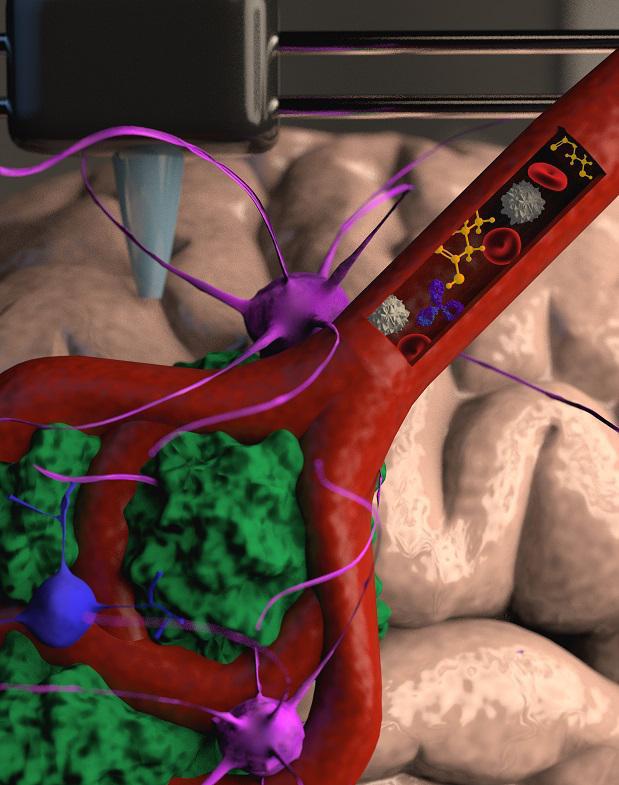
Illustration.
Credit: Veronica Hughes, PhD of STEAM visuals

The study was led by Prof. Ronit Satchi-Fainaro, Sackler Faculty of Medicine and Sagol School of Neuroscience, Director of the Cancer Biology Research Center, Head of the Cancer Research and Nanomedicine Laboratory and Director of the Morris Kahn 3D-BioPrinting for Cancer Research Initiative, at Tel Aviv University.

The new technology was developed by PhD student Lena Neufeld, together with other researchers at Prof. Satchi-Fainaro’s laboratory:  Eilam Yeini, Noa Reisman, Yael Shtilerman, Dr. Dikla Ben-Shushan, Sabina Pozzi, Dr. Galia Tiram, Dr. Anat Eldar-Boock and Dr. Shiran Farber.  

The 3D-bioprinted models are based on samples from patients, taken directly from operating rooms at the Tel Aviv Sourasky Medical Center. The new study’s results were published today in the prestigious journal Science Advances.

“Glioblastoma is the most lethal cancer of the central nervous system, accounting for most brain malignancies”

“Glioblastoma is the most lethal cancer of the central nervous system, accounting for most brain malignancies,” says Prof. Satchi-Fainaro. “In a previous study, we identified a protein called P-Selectin, produced when glioblastoma cancer cells encounter microglia – cells of the brain’s immune system. We found that this protein is responsible for a failure in the microglia, causing them to support rather than attack the deadly cancer cells, helping the cancer spread. However, we identified the protein in tumors removed during surgery, but not in glioblastoma cells grown on 2D plastic petri dishes in our lab. The reason is that cancer, like all tissues, behaves very differently on a plastic surface than it does in the human body. Approximately 90% of all experimental drugs fail at the clinical stage because the success achieved in the lab is not reproduced in patients.”
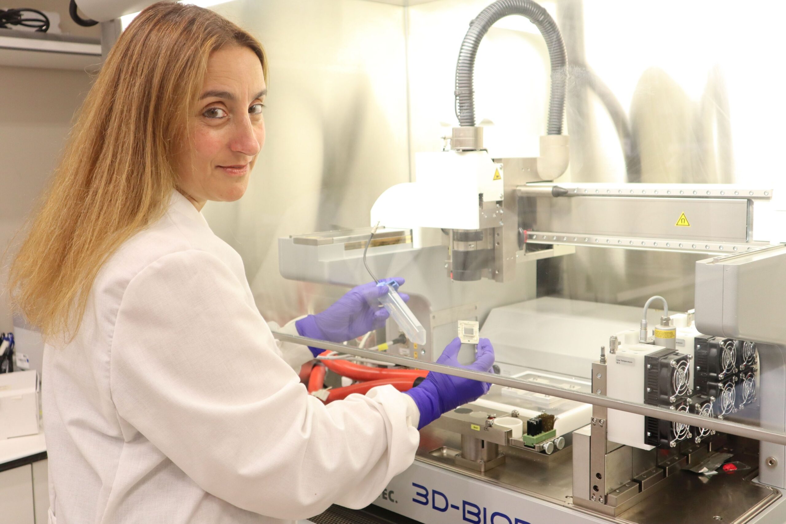
Prof. Ronit Satchi-Fainaro

To address this problem, the research team led by Prof. Satchi-Fainaro and PhD student Lena Neufeld, recipient of the prestigious Dan David Fellowship, created the first 3D-bioprinted model of a glioblastoma tumor, which includes 3D cancer tissue surrounded by extracellular matrix, which communicates with its microenvironment via functional blood vessels.
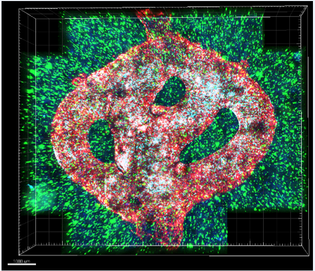
Microscopic image of the 3D-bioprinted glioblastoma model. The bioprinted blood vessels are covered with endothelial cells (red) and pericytes (cyan). The blood vessels are surrounded with a brain-mimicking tissue composed of gliblastoma cells (blue) and the brain microenvironment cells (green). Different drugs or cells can be perfused through the 3D-bioprinted blood vessels to test their effect on the tumor tissue
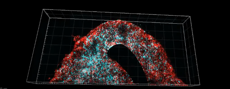
“It’s not only the cancer cells…”

“It’s not only the cancer cells,” explains Prof. Satchi-Fainaro. “It’s also the cells of the microenvironment in the brain; the astrocytes, microglia and blood vessels connected to a microfluidic system – namely a system enabling us to deliver substances like blood cells and drugs to the tumor replica. Each model is printed in a bioreactor we have designed in the lab, using a hydrogel sampled and reproduced from the extracellular matrix taken from the patient, thereby simulating the tissue itself. The physical and mechanical properties of the brain are different from those of other organs, like the skin, breast, or bone. Breast tissue consists mostly of fat, bone tissue is mostly calcium; each tissue has its own properties, which affect the behavior of cancer cells and how they respond to medications. Growing all types of cancer on identical plastic surfaces is not an optimal simulation of the clinical setting.”

After successfully printing the 3D tumor, Prof. Satchi-Fainaro and her colleagues demonstrated that unlike cancer cells growing on petri dishes, the 3D-bioprinted model has the potential to be effective for rapid, robust, and reproducible prediction of the most suitable treatment for a specific patient.

“We proved that our 3D model is better suited for prediction of treatment efficacy, target discovery and drug development in three different ways.

First, we tested a substance that inhibited the protein we had recently discovered, P-Selectin, in glioblastoma cell cultures grown on 2D petri dishes, and found no difference in cell division and migration between the treated cells and the control cells which received no treatment. In contrast, in both animal models and in the 3D-bioprinted models, we were able to delay the growth and invasion of glioblastoma by blocking the P-Selectin protein.

This experiment showed us why potentially effective drugs rarely reach the clinic simply because they fail tests in 2D models, and vice versa: why drugs considered a phenomenal success in the lab, ultimately fail in clinical trials. In addition, collaborating with the lab of Dr. Asaf Madi of the Department of Pathology at TAU’s Faculty of Medicine, we conducted genetic sequencing of the cancer cells grown in the 3D-bioprinted model, and compared them to both cancer cells grown on 2D plastic and cancer cells taken from patients.

Thus, we demonstrated a much greater resemblance between the 3D-bioprinted tumors and patient-derived glioblastoma cells grown together with brain stromal cells in their natural environment. Through time, the cancer cells grown on plastic changed considerably, finally losing any resemblance to the cancer cells in the patient’s brain tumor sample.

The third proof was obtained by measuring the tumor growth rate. Glioblastoma is an aggressive disease partially because it is unpredictable: when the heterogeneous cancer cells are injected separately into model animals, the cancer will remain dormant in some, while in others, an active tumor will develop rapidly. This makes sense because we, as humans, can die peacefully of old age without ever knowing we have harbored such dormant tumors. On the dish in the lab, however, all tumors grow at the same rate and spread in the same rate. In our 3D-bioprinted tumor, the heterogeneity is maintained and development is similar to the broad spectrum that we see in patients or animal models.”

“…perhaps the most exciting aspect is finding novel druggable target proteins and genes in cancer cells…”

According to Prof. Satchi-Fainaro, this innovative approach will also enable the development of new drugs, as well as discovery of new drug targets – at a much faster rate than today. Hopefully, in the future, this technology will facilitate personalized medicine for patients.

“If we take a sample from a patient’s tissue, together with its extracellular matrix, we can 3D-bioprint from this sample 100 tiny tumors and test many different drugs in various combinations to discover the optimal treatment for this specific tumor. Alternately, we can test numerous compounds on a 3D-bioprinted tumor and decide which is most promising for further development and investment as a potential drug.

But perhaps the most exciting aspect is finding novel druggable target proteins and genes in cancer cells – a very difficult task when the tumor is inside the brain of a human patient or model animal. Our innovation gives us unprecedented access, with no time limits, to 3D tumors mimicking better the clinical scenario, enabling optimal investigation.”
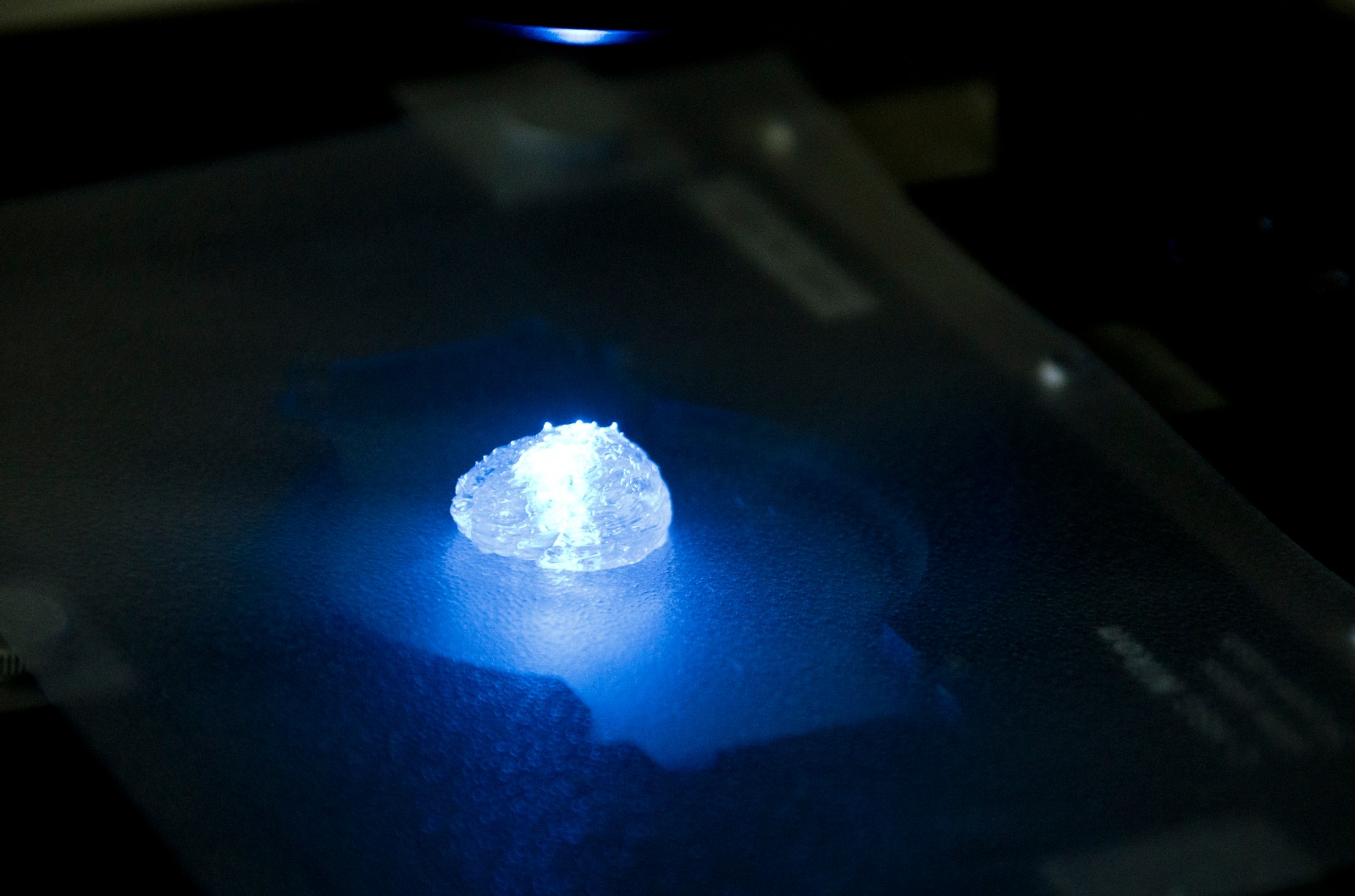
Illustration for demonstration of 3D printing of a tumor in a brain Microenvironment according to a computed 3D model

The study was funded by the Morris Kahn Foundation, European Research Council (ERC), Israel Cancer Research Fund (ICRF), the Israel Cancer Association and Israel Science Foundation (ISF), and Check Point Software Technologies LTD.

Continue reading

New Warning Sign for Breast Cancer

TAU-led research lays groundwork for preventive treatment that may save millions of lives.

A team led by Tel Aviv University identified a new indicator of metastatic breast cancer, laying the groundwork for preventive treatment that could save millions of lives.

Metastatic breast cancer, also known as Stage 4 breast cancer, occurs when cancer has spread, or “metastasized,” to other parts of the body. Mortality from breast cancer is almost exclusively a result of tumor metastasis, and lungs are one of the main metastatic sites. The five-year survival rate for women with metastatic breast cancer is estimated at 28%.

Investigating The “Black Box” of Breast Cancer 

“Breast cancer patients, as well as patients with many other types of cancer, do not die from the primary tumor, but from distant metastases which have developed, sometimes after years, in essential organs such as the lungs and brain,” said the study’s lead researcher, Prof. Neta Erez, Chair of the Department of Pathology at TAU’s Sackler Faculty of Medicine. “Understanding the body’s preparation for the reception of metastases at an early stage may save millions of lives.”

The researchers explain that metastases can appear several years after the initial cases are treated. Today, methods used for follow-up screening identify metastases only when they are quite large–when the disease is at an advanced stage and unlikely to be cured.

For this reason, Erez’s research group is investigating the black box—the time period between apparent recovery and the appearance of metastases to understand the metastatic process and to find ways of blocking it in early stages. Their research in recent years has revealed that certain tissues, in organs where the metastases are set to arrive, “prepare the area” for reception and produce a hospitable environment for them, a long time before the appearance of the metastases themselves. In the present study, the research team searched for signs of these changes, which may be used in the future to identify the start of the process that predicts metastases. The researchers identified these changes in the area known as “the micro-environment” of the tumor, and specifically in connective tissue known as fibroblasts which are found in the lungs among other places. 

“In a normal situation, fibroblasts play a central role in healing wounds and injury to the lungs, but recent studies revealed that cancer is successful in recruiting them and causing them to produce a supportive environment for it,” said Erez.

What is Happening in the Micro-environment of the Metastases?

The researchers compared genes sequenced from healthy lungs, from lungs with micro-metastases (very small metastases which cannot be identified using existing clinical tools) and from lungs with large metastases, in a state of advanced disease.

By identifying and comparing the respective development in the three different types of sample tissues, the researchers succeeded, for the first time, in characterizing the process that occurs in the micro-environment of the metastases. The findings provide valuable understanding about how cancer cells grow, which can then be leveraged for detection by existing imaging methods and treated to prevent metastasis.

The study’s leading research team from Erez’s laboratory included Dr. Ophir Shani and Dr. Yael Raz along with additional researchers from Tel Aviv University, Sheba Medical Center at Tel HaShomer, Tel Aviv Sourasky Medical Center (Ichilov Hospital), and the Weizmann Institute of Science. The findings were published in the prestigious peer-reviewed journal eLife.  

Featured image: Prof. Neta Erez (Photo: Michal Kidron)

COVID-19 Immunity Varies Among Genders and Age Groups

TAU researchers contribute a new piece to the puzzle on the effectiveness of COVID-19 vaccination.

As experts continue to learn more about immune responses to COVID-19 and the effectiveness of vaccines, researchers from Tel Aviv University have contributed a new piece to the puzzle. A joint study conducted by researchers from TAU and the Shamir Medical Center (Assaf Harofe) indicates that the level of antibodies changes according to age groups, gender, symptoms, and time elapsed since vaccination. The findings are the latest from the researchers in a series of studies aimed at providing reliable measures on the effectiveness of COVID-19 vaccination.

The new study examined the level of antibodies in over 26,000 blood samples taken from COVID-19 convalescents, as well as vaccinated and unvaccinated individuals.

In vaccinated individuals, the researchers found differences between women and men in the concentration of antibodies in the blood relative to both age and gender. In women, the level of antibodies begins to rise from the age of 51, and is higher than the levels found in men of similar age. This phenomenon may be related change in levels of the estrogen hormone, observed around this age, which affects the immune system. In men, a rise in antibody levels is seen at an earlier age, starting around 35, and may be related to changes in levels of testosterone and the effect on the immune system.

In young adults, a high concentration of antibodies generally signals a strong healthy functioning immune response, while in older demographics it typically indicates overreaction of the immune system associated with severe illness. In general, young adults were found to have a higher level of antibodies sustained for a longer period of time compared to older vaccinated persons. The findings further validate existing evidence that, depending on age, higher antibody count isn’t necessarily equivalent to higher rates of recovery.

Furthermore, the study found that the immune response of vaccinated individuals (after two doses) is much stronger than that of people who have recovered from COVID-19. The findings show that vaccinated individuals have four times the level of antibodies compared to convalescents.

The study was conducted by Tel Aviv University’s Prof. Noam Shomron, Head of the Computational Genomics Laboratory at the Sackler Faculty of Medicine and a member of the Edmond J. Safra Center for Bioinformatics and Dr. Adina Bar Chaim from the Shamir Medical Center. The data were collected by Dr. Ramzia Abu Hamad from the Shamir Medical Center, and analysis was conducted by Guy Shapira, a PhD student at Prof. Shomron’s laboratory. The study was published in Medrxiv

New study found differences between women and men in the level of COVID-19 antibodies

Prof. Noam Shomron, Head of the Computational Genomics Laboratory at the Sackler Faculty of Medicine and a member of the Edmond J. Safra Center for Bioinformatics

A joint study conducted by researchers from Tel Aviv University and the Shamir Medical Center (Asaf Harofe) examined the level of antibodies in over 26,000 blood samples taken from COVID-19 convalescents, as well as vaccinated and unvaccinated individuals. The serological results indicate that the level of antibodies changes according to age groups, gender, symptoms, and time elapsed since vaccination. The study was published in Medrxiv.

A difference was found between vaccinated women and men, in the concentration of antibodies in the blood relative to both age and gender. In women, the level of antibodies begins to rise from the age of 51, and is higher than the levels found in men of similar age. This phenomenon may be related change in levels of the estrogen hormone, observed around this age, which affects the immune system. In men, a rise in antibody levels is seen at an earlier age, starting around 35. This may be related to changes in levels of the male sex hormone testosterone, and the effect on the immune system.

In young adults, a high concentration of antibodies is usually the result of a strong immune response, while in older people it typically indicates overreaction of the immune system associated with severe illness.

Dr. Adina Bar Chaim from the Shamir Medical Center

Main trends and findings:

  1. The immune response of individuals who have received two doses of the vaccine is much stronger than that of people who have recovered from COVID-19. In fact, the level of antibodies found in the blood of vaccinated persons was 4 times higher than that found in convalescents.
  2. A difference was found between convalescent males and females – in antibody concentration in the blood relative to both age and gender. In women, the concentration begins to rise from the age of 51, and it is higher than the levels found in men of similar age. This phenomenon may be related to the change in levels of the estrogen hormone, observed around this age, which affects the immune system. In men, a rise in antibody levels is seen at an earlier age, starting around 35. This may be related to changes in levels of the male sex hormone testosterone, and its effect on the immune system.

In young adults, a high concentration of antibodies is usually the result of a strong immune response, while in older people it usually indicates overreaction of the immune system associated with severe illness.

  1. In general, young adults were found to have a higher level of antibodies sustained for a longer period of time compared to older vaccinated persons. A decrease of tens of percent was observed over time between the younger and very old age groups.

Conclusion: Further research is required in order to obtain an in-depth understanding of the immune system’s response to COVID-19, to recovery from the disease, and to the vaccine. We hope that in the future we will be able to supply a reliable measure for the effectiveness of vaccination, correlated with age, gender and symptoms.

The study was conducted by Tel Aviv University’s Prof. Noam Shomron, Head of the Computational Genomics Laboratory at the Sackler Faculty of Medicine and a member of the Edmond J. Safra Center for Bioinformatics and Dr. Adina Bar Chaim from the Shamir Medical Center. The data were collected by Dr. Ramzia Abu Hamad from the Shamir Medical Center, and analysis was conducted by Guy Shapira, a PhD student at Prof. Shomron’s laboratory.

Continue reading

A world first: Technology that restores the sense of touch in nerves damaged as a result of amputation or injury

Cut your finger and lost your sense of touch? There’s hope yet.

  • Researchers have developed a sensor that can be implanted anywhere in the body, for example under the tip of a severed finger; the sensor connects to another nerve that functions properly and restores tactile sensation to the injured nerve.
  • This unique development is biocompatible (“human-body friendly”) and does not require electricity, wires, or batteries.

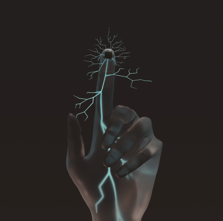
Tel Aviv University’s new and groundbreaking technology inspires hope among people who have lost their sense of touch in the nerves of a limb following amputation or injury. The technology involves a tiny sensor that is implanted in the nerve of the injured limb, for example in the finger, and is connected directly to a healthy nerve. Each time the limb touches an object, the sensor is activated and conducts an electric current to the functioning nerve, which recreates the feeling of touch. The researchers emphasize that this is a tested and safe technology that is suited to the human body and could be implanted anywhere inside of it once clinical trials will be done.

The technology was developed under the leadership of a team of experts from Tel Aviv University: Dr. Ben M. Maoz, Iftach Shlomy, Shay Divald, and Dr. Yael Leichtmann-Bardoogo from the Department of Biomedical Engineering, Fleischman Faculty of Engineering, in collaboration with Keshet Tadmor from the Sagol School of Neuroscience and Dr. Amir Arami from the Sackler School of Medicine and the Microsurgery Unit in the Department of Hand Surgery at Sheba Medical Center. The study was published in the prestigious journal ACS Nano.

The researchers say that this unique project began with a meeting between the two Tel Aviv University colleagues – biomedical engineer Dr. Maoz and surgeon Dr. Arami. “We were talking about the challenges we face in our work,” says Dr. Maoz, “and Dr. Arami shared with me the difficulty he experiences in treating people who have lost tactile sensation in one organ or another as a result of injury. It should be understood that this loss of sensation can result from a very wide range of injuries, from minor wounds – like someone chopping a salad and accidentally cutting himself with the knife – to very serious injuries. Even if the wound can be healed and the injured nerve can be sutured, in many cases the sense of touch remains damaged. We decided to tackle this challenge together, and find a solution that will restore tactile sensation to those who have lost it.”

In recent years, the field of neural prostheses has made promising developments to improve the lives of those who have lost sensation in their limbs by implanting sensors in place of the damaged nerves. But the existing technology has a number of significant drawbacks, such as complex manufacturing and use, as well as the need for an external power source, such as a battery. Now, the researchers at Tel Aviv University have used state-of-the-art technology called a triboelectric nanogenerator (TENG) to engineer and test on animal models a tiny sensor that restores tactile sensation via an electric current that comes directly from a healthy nerve and doesn’t require a complex implantation process or charging.

The researchers developed a sensor that can be implanted on a damaged nerve under the tip of the finger; the sensor connects to another nerve that functions properly and restores some of the tactile sensation to the finger. This unique development does not require an external power source such as electricity or batteries. The researchers explain that the sensor actually works on frictional force: whenever the device senses friction, it charges itself.

The device consists of two tiny plates less than half a centimeter by half a centimeter in size. When these plates come into contact with each other, they release an electric charge that is transmitted to the undamaged nerve. When the injured finger touches something, the touch releases tension corresponding to the pressure applied to the device – weak tension for a weak touch and strong tension for a strong touch – just like in a normal sense of touch.

The researchers explain that the device can be implanted anywhere in the body where tactile sensation needs to be restored, and that it actually bypasses the damaged sensory organs. Moreover, the device is made from biocompatible material that is safe for use in the human body, it does not require maintenance, the implantation is simple, and the device itself is not externally visible.

According to Dr. Maoz, after testing the new sensor in the lab (with more than half a million finger taps using the device), the researchers implanted it in the feet of the animal models. The animals walked normally, without having experienced any damage to their motor nerves, and the tests showed that the sensor allowed them to respond to sensory stimuli. “We tested our device on animal models, and the results were very encouraging,” concludes Dr. Maoz. “Next, we want to test the implant on larger models, and at a later stage implant our sensors in the fingers of people who have lost the ability to sense touch. Restoring this ability can significantly improve people’s functioning and quality of life, and more importantly, protect them from danger. People lacking tactile sensation cannot feel if their finger is being crushed, burned or frozen.”

Dr. Maoz’s laboratory:

https://www.maozlab.com/

  The article:

https://pubs.acs.org/doi/full/10.1021/acsnano.0c10141


Continue reading

New nanotech from TAU produces “healthy” electric current from the human body itself

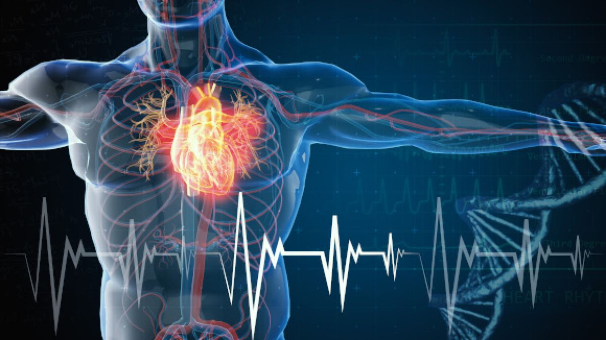
Approach allows for the charging of cardiac pacemakers using only the heartbeat, eliminating the need for batteries

A new nanotechnology development from an international research team led by Tel Aviv University researchers will make it possible to generate electric currents and voltage within the human body itself through the activation of various organs using mechanical force. The development involves a new and very strong biological material, similar to collagen, which is non-toxic and causes no harm to the body’s tissues.

The researchers believe that this new nanotechnology has many potential applications in medicine, including harvesting clean energy to operate pacemakers and other devices implanted in the body through the body’s natural movements, eliminating the need for batteries and the surgery required to replace them.

The study was led by Professor Ehud Gazit of TAU’s Shmunis School of Biomedicine and Cancer Research at the George S. Wise Faculty of Life Sciences, the Department of Materials Science and Engineering at the Fleischman Faculty of Engineering and the Center for Nanoscience and Nanotechnology, along with his lab team, Dr. Santu Bera and Dr. Wei Ji.

Researchers from the Weizmann Institute and a number of research institutes in Ireland, China and Australia also took part in the study, which was published in Nature Communications.

“Collagen is the most prevalent protein in the human body, constituting about 30% of all of the proteins in our body,” Professor Gazit, who is also Founding Director of TAU’s Blavatnik Center for Drug Discovery, explains. “It is a biological material with a helical structure and a variety of important physical properties, such as mechanical strength and flexibility, which are useful in many applications. However, because the collagen molecule itself is large and complex, researchers have long been looking for a minimalistic, short and simple molecule that is based on collagen and exhibits similar properties.

“About a year and a half ago our group published a study in which we used nanotechnological means to engineer a new biological material that meets these requirements,” Professor Gazit continues. “It is a tripeptide — a very short molecule called Hyp-Phe-Phe consisting of only three amino acids — capable of a simple process of self-assembly of forming a collagen-like helical structure that is flexible and boasts a strength similar to that of the metal titanium.

“In the present study, we sought to examine whether the new material we developed bears piezoelectricity, another feature that characterizes collagen. Piezoelectricity is the ability of a material to generate electric currents and voltage as a result of the application of mechanical force, or vice versa, to create a mechanical force as the result of exposure to an electric field.”

The researchers created nanometric structures of the engineered material, and with the help of advanced nanotechnology tools applied mechanical pressure on them. The experiment revealed that the material does indeed produce electric currents and voltage as a result of the pressure.

Moreover, tiny structures of mere hundreds of nanometers demonstrated one of the highest levels of piezoelectric ability ever discovered, comparable or superior to that of the piezoelectric materials commonly found in today’s market, most of which contain lead and are unsuitable for medical applications.

According to the researchers, the discovery of piezoelectricity of this magnitude in a nanometric material is of great significance, as it demonstrates the ability of the engineered material to serve as a kind of tiny motor for very small devices. Next, the researchers plan to apply crystallography and computational quantum mechanical methods (density functional theory) in order to gain an in-depth understanding of the material’s piezoelectric behavior and thereby enable the accurate engineering of crystals for the building of biomedical devices.

“Most of the piezoelectric materials that we know of today are toxic lead-based materials, or polymers, meaning they are not environmentally and human body-friendly,” Professor Gazit says. “Our new material, however, is completely biological and suitable for uses within the body.

“For example, a device made from this material may replace a battery that supplies energy to implants like pacemakers, though it should be replaced from time to time. Body movements like heartbeats, jaw movements, bowel movements, or any other movement that occurs in the body on a regular basis will charge the device with electricity, which will continuously activate the implant.”

His current focus is on the development of medical devices, but Professor Gazit emphasizes that “environmentally friendly piezoelectric materials, such as the one we have developed, have tremendous potential in a wide range of areas because they produce green energy using mechanical force that is being used anyway. For example, a car driving down the street can turn on the streetlights. These materials may also replace lead-containing piezoelectric materials that are currently in widespread use, but that raise concerns about the leakage of toxic metal into the environment.”

Introducing the world’s thinnest technology – only two atoms thick

Technological breakthrough from Tel Aviv University

The research team
  • The new technology, enabling the storage of information in the thinnest unit known to science, is expected to improve future electronic devices in terms of density, speed, and efficiency.

  • The allowed quantum-mechanical electron tunneling through the atomically thin film may boost the information reading process much beyond current technologies.

  • The technology involves laterally sliding one-atom-thick layers of boron and nitrogen one over the other – a new way to switch electric polarization on/off.

A scientific breakthrough: Researchers from Tel Aviv University have engineered the world’s tiniest technology, with a thickness of only two atoms. According to the researchers, the new technology proposes a way for storing electric information in the thinnest unit known to science, in one of the most stable and inert materials in nature. The allowed quantum-mechanical electron tunneling through the atomically thin film may boost the information reading process much beyond current technologies.

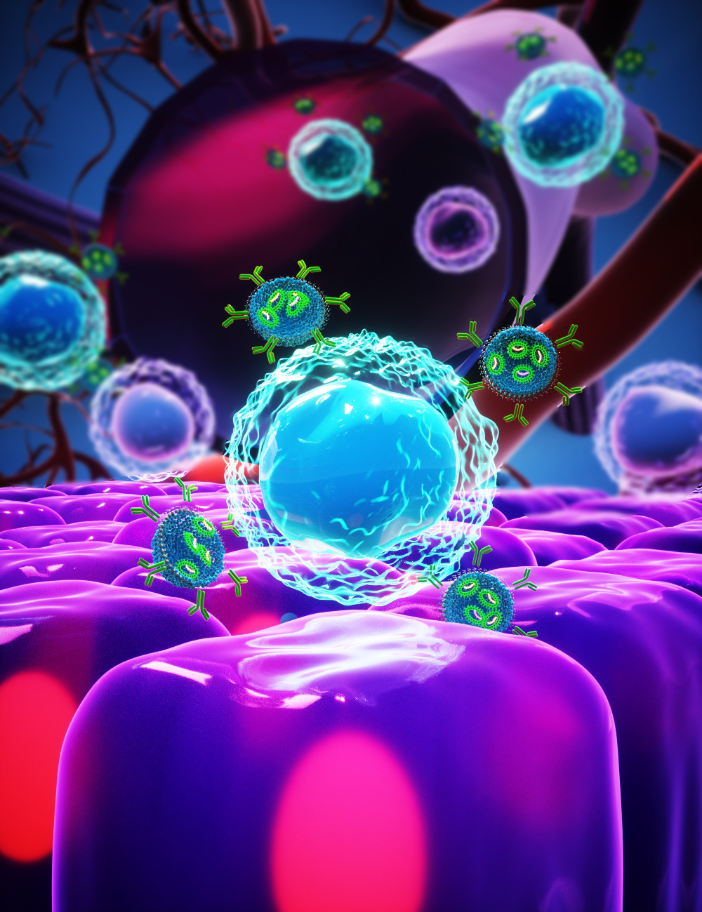
The research was performed by scientists from the Raymond and Beverly Sackler School of Physics and Astronomy and Raymond and Beverly Sackler School of Chemistry.  The group includes Maayan Vizner Stern, Yuval Waschitz, Dr. Wei Cao, Dr. Iftach Nevo, Prof. Eran Sela, Prof. Michael Urbakh, Prof. Oded Hod, and Dr. Moshe Ben Shalom. The work is now published in Science magazine.

“Our research stems from curiosity about the behavior of atoms and electrons in solid materials, which has generated many of the technologies supporting our modern way of life,” says Dr. Ben Shalom. “We (and many other scientists) try to understand, predict, and even control the fascinating properties of these particles as they condense into an ordered structure that we call a crystal. At the heart of the computer, for example, lies a tiny crystalline device designed to switch between two states indicating   different responses – “yes” or “no”, “up” or “down” etc. Without this dichotomy – it is not possible to encode and process information. The practical challenge is to find a mechanism that would enable switching in a small, fast, and inexpensive device.

Current state-of-the-art devices consist of tiny crystals that contain only about a million atoms (about a hundred atoms in height, width, and thickness) so that a million of these devices can be squeezed about a million times into the area of one coin, with each device switching at a speed of about a million times per second.

Following the technological breakthrough, the researchers were able, for the first time, to reduce the thickness of the crystalline devices to two atoms only. Dr. Ben Shalom emphasizes that such a thin structure enables memories based on the quantum ability of electrons to hop quickly and efficiently through barriers that are just several atoms thick. Thus, it may significantly improve electronic devices in terms of speed, density, and energy consumption.

In the study, the researchers used a two-dimensional material: one-atom-thick layers of boron and nitrogen, arranged in a repetitive hexagonal structure. In their experiment, they were able to break the symmetry of this crystal by artificially assembling two such layers. “In its natural three-dimensional state, this material is made up of a large number of layers placed on top of each other, with each layer rotated 180 degrees relative to its neighbors (antiparallel configuration)” says Dr. Ben Shalom. “In the lab, we were able to artificially stack the layers in a parallel configuration with no rotation, which hypothetically places atoms of the same kind in perfect overlap despite the strong repulsive force between them (resulting from their identical charges). In actual fact, however, the crystal prefers to slide one layer slightly in relation to the other, so that only half of each layer’s atoms are in perfect overlap, and those that do overlap are of opposite charges – while all others are located above or below an empty space – the center of the hexagon. In this artificial stacking configuration the layers are quite distinct from one another. For example, if in the top layer only the boron atoms overlap, in the bottom layer it’s the other way around.”

Dr. Ben Shalom also highlights the work of the theory team, who conducted numerous computer simulations “Together we established deep understanding of why the system’s electrons arrange themselves just as we had measured in the lab. Thanks to this fundamental understanding, we expect fascinating responses in other symmetry-broken layered systems as well,” he says.

Maayan Wizner Stern, the PhD student who led the study, explains: “The symmetry breaking we created in the laboratory, which does not exist in the natural crystal, forces the electric charge to reorganize itself between the layers and generate a tiny internal electrical polarization perpendicular to the layer plane. When we apply an external electric field in the opposite direction the system slides laterally to switch the polarization orientation. The switched polarization remains stable even when the external field is shut down. In this the system is similar to thick three-dimensional ferroelectric systems, which are widely used in technology today.”

“The ability to force a crystalline and electronic arrangement in such a thin system, with unique polarization and inversion properties resulting from the weak Van der Waals forces between the layers, is not limited to the boron and nitrogen crystal,” adds Dr. Ben Shalom. “We expect the same behaviors in many layered crystals with the right symmetry properties. The concept of interlayer sliding as an original and efficient way to control advanced electronic devices is very promising, and we have named it Slide-Tronics”.

Maayan Vizner Stern concludes: “We are excited about discovering what can happen in other states we force upon nature and predict that other structures that couple additional degrees of freedom are possible. We hope that miniaturization and flipping through sliding will improve today’s electronic devices, and moreover, allow other original ways of controlling information in future devices. In addition to computer devices, we expect that this technology will contribute to detectors, energy storage and conversion, interaction with light, etc. Our challenge, as we see it, is to discover more crystals with new and slippery degrees of freedom.”

The study was funded through support from the European Research Council (ERC starting grant), the Israel Science Foundation (ISF), and the Ministry of Science and Technology (MOST).

Continue reading

Want to Live a Long Life? Consider Investing in Your Marriage.

TAU researchers find link between marriage quality and life expectancy.

Want to live a long and healthy life? For the men among us, TAU researchers’ best advice is to invest in our relationship.

As Harmful as Smoking

“Our study shows that the quality of marriage and family life has health implications for life expectancy. Men who reported they perceived their marriage as failure died younger than those who experienced their marriages as very successful. In other words, the level of satisfaction with marriage has emerged as a predictive factor for life expectancy at a rate comparable with smoking (smokers versus non-smokers) and physical activity (activity versus inactivity)”, said one of the study’s lead researchers, Dr. Shahar Lev-Ari, head of the Department of Health Promotion at TAU’s School of Public Health, Sackler Faculty of Medicine.

“Furthermore, it’s important to note that we observed a higher risk among relatively young men, under the age of 50. At a higher age, the gap is smaller, perhaps due to processes of adjustment that life partners go through over time.”

The study was based on extensive health data from more than 30 years of research that tracked the deaths of 10,000 Israeli men.

In addition to Dr. Lev-Ari, lead researchers from the School of Public Health at the Sackler Faculty of Medicine also included: Prof. Uri Goldbort from the Department of Epidemiology and Preventive Medicine, who initiated and managed the long-term study, and Dr. Yiftah Gapner, from the Department of Epidemiology and Preventive Medicine. The article was published in the Journal of Clinical Medicine.

As part of the study, the researchers conducted statistical analyses of a database launched in the 1960s. For 32 years, they tracked the health and behavior of 10,000 male Israel state employees, paying special attention to death from strokes and premature death in general.

At the beginning of the study, most of the participants were in their 40s. Since then, 64% died from a range of illnesses. “We wanted to analyze the data gathered longitudinally using various parameters to identify behavioral and psychosocial risk factors that can predict death from a CVA [a cerebrovascular accident or, in other words, a stroke] and premature death for any reason,” Dr. Lev-Ari explains.

Early in the 32-year-long study, participants were asked to rank their level of marriage satisfaction on a scale of 1 (marriage is very successful) to 4 (marriage is unsuccessful). To the researchers’ surprise, this scale would prove to be a predictive factor for life expectancy, highly similar to smoking and lack of physical activity. The number of deceased from a stroke was 69% higher among those who ranked their marriage satisfaction at 4 (i.e. marriage is unsuccessful) compared to those who ranked their marriage satisfaction very highly. The overall mortality was 19% higher among the unhappily married. The researchers note that the gaps were even larger among men who were relatively young (under 50) at the beginning of the study.

In addition, the researchers conducted a statistical analysis of all known risk factors contributing to death from cardiovascular diseases, such as diabetes, hypertension, excessive BMI, and socioeconomic status. Here, too, the data showed that the relative risk of death for any reason among the unhappily married was 1.21 higher than among those satisfied with their marriages. This rate is similar to data cited in medical literature regarding smokers and those leading a sedentary life.

Your list of healthy habits just got a bit longer, guys. But remember, knowledge is power – and next time you go to the gym, perhaps you could make it a date?

A world first: Targeted delivery of therapeutic RNAs only to cancer cells, with no harm caused to healthy cells

Tel Aviv University’s Groundbreaking Technology:

The “door-to-door service” that delivers therapeutic RNA payloads directly to cancer cells and other diseased cells 

  • The groundbreaking technology may revolutionize the treatment of cancer and a wide range of diseases and medical conditions.

  • Researcher Prof. Peer: “Our development actually changes the world of therapeutic antibodies. Today we flood the body with antibodies that, although selective, also damage healthy cells. We have now removed the uninfected cells from the equation, and, via a simple injection, succeeded in targeting only the cells that are inflamed at that given moment.”

  • The study was published in the prestigious scientific journal Nature.

Tel Aviv University’s groundbreaking technology may revolutionize the treatment of cancer and a wide range of diseases and medical conditions. In the framework of this study, the researchers were able to create a new method of transporting RNA-based drugs to a subpopulation of immune cells involved in the inflammation process, and target the disease-inflamed cell without causing damage to other cells.

The study was led by Prof. Dan Peer, a global pioneer in the development of RNA-based therapeutic delivery. He is Tel Aviv University’s Vice President for Research and Development, head of the Center for Translational Medicine and a member of both the Shmunis School of Biomedicine and Cancer Research, George S. Wise Faculty of Life Sciences, and the Center for Nanoscience and Nanotechnology. The study was published in the prestigious scientific journal Nature.

Prof. Peer: “Our development actually changes the world of therapeutic antibodies. Today we flood the body with antibodies that, although selective, damage all the cells that express a specific receptor, regardless of their current form. We have now taken out of the equation healthy cells that can help us, that is, uninflamed cells, and via a simple injection into the bloodstream can silence, express or edit a particular gene exclusively in the cells that are inflamed at that given moment.”

As part of the study, Prof. Peer and his team were able to demonstrate this groundbreaking development in animal models of inflammatory bowel diseases such as Crohn’s disease and colitis, and improve all inflammatory symptoms, without performing any manipulation on about 85% of the immune system cells. Behind the innovative development stands a simple concept, targeting to a specific receptor conformation.

“On every cell envelope in the body, that is, on the cell membrane, there are receptors that select which substances enter the cell,” explains Prof. Peer. “If we want to inject a drug, we have to adapt it to the specific receptors on the target cells, otherwise it will circulate in the bloodstream and do nothing. But some of these receptors are dynamic – they change shape on the membrane according to external or internal signals. We are the first in the world to succeed in creating a drug delivery system that knows how to bind to receptors only in a certain situation, and to skip over the other identical cells, that is, to deliver the drug exclusively to cells that are currently relevant to the disease.”

Previously, Prof. Peer and his team developed delivery systems based on fatty nanoparticles – the most advanced system of its kind; this system has already received clinical approval for the delivery of RNA-based drugs to cells. Now, they are trying to make the delivery system even more selective.

According to Prof. Peer, the new breakthrough has possible implications for a wide range of diseases and medical conditions. “Our development has implications for many types of blood cancers and various types of solid cancers, different inflammatory diseases, and viral diseases such as the coronavirus. We now know how to wrap RNA in fat-based particles so that it binds to specific receptors on target cells,” he says. “But the target cells are constantly changing. They switch from ‘binding’ to ‘non-binding’ mode in accordance with the circumstances. If we get a cut, for example, not all of our immune system cells go into a ‘binding’ state, because we do not need them all in order to treat a small incision. That is why we have developed a unified protein that knows how to bind only to the active state of the receptors of the immune system cells. We tested the protein we developed in animal models of inflammatory bowel disease, both acute and chronic.”

Prof. Peer adds, “We were able to organize the delivery system in such a way that we target to only 14.9% of the cells that were involved in the inflammatory condition of the disease, without adversely affecting the other, non-involved, cells, which are actually completely healthy cells. Through specific binding to the cell sub-population, while delivering the RNA payload we were able to improve all indices of inflammation, from the animal’s weight to pro-inflammatory cytokines. We compared our results with those of antibodies that are currently on the market for Crohn’s and colitis patients, and found that our results were the same or better, without causing most of the side effects that accompany the introduction of antibodies into the entire cell population. In other words, we were able to deliver the drug ‘door-to-door,’ directly to the diseased cells.”

The study was led by Prof. Peer, together with Dr. Niels Dammes, a postdoctoral fellow from the Netherlands, with the collaboration of Dr. Srinivas Ramishetti, Dr. Meir Goldsmith and Dr. Nuphar Veiga, from Prof. Dan Peer’s lab. Professors Jason Darling and Alan Packard of Harvard University in the United States also participated. The study was funded by the European Union, in the framework of the European Research Council (ERC).

Continue reading

Combating Antibiotic Resistance

Discovery may contribute to new treatments for infectious diseases.

A new TAU study revealed a mechanism through which “good” viruses can attack the systems of “bad” bacteria, destroy them and block their reproduction.

“Good” Viruses Kill “Bad” Bacteria

The researchers demonstrated that the “good” virus (bacteriophage) is able to block the replication mechanism of the bacteria’s DNA without damaging its own, noting that the ability to distinguish between oneself and others is crucial in nature. The discovery reveals one more fascinating aspect of the mutual relations between bacteria and bacteriophages and may lead to a better understanding of bacterial mechanisms for evading bacteriophages, as well as ways for using bacteriophages to combat bacteria. The study, published recently in PNAS – Proceedings of the National Academy of Sciences, was led by Prof. Udi Qimron, Dr. Dor Salomon, Dr. Tridib Mahata and Shahar Molshanski-Mor of the Sackler Faculty of Medicine. Other participants included Prof. Tal Pupko, Head of The Shmunis School of Biomedicine and Cancer Research and also of TAU’s new AI and Data Science Center; Dr. Oren Avram of The George S. Wise Faculty of Life Sciences; and Dr. Ido Yosef, Dr. Moran Goren, Dr. Miriam Kohen-Manor and Dr. Biswanath Jana of the Sackler Faculty of Medicine.

A Great Scientific Challenge

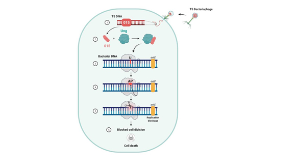
Prof. Qimron explains that the antibiotic resistance of bacteria is one of the greatest challenges faced by scientists today. One potential solution may lie in further investigation of the targeted eradication of bacteria by “good” bacteriophages; namely, understanding bacteriophage mechanisms for taking over bacteria as a basis for the development of new tools to combat bacterial pathogens. With this solution in mind, the current study unveiled the mechanism by which the bacteriophage takes control of the bacteria. The researchers found that a bacteriophage protein uses a DNA-repair protein in the bacteria to “cunningly” cut the bacteria’s DNA as it is being repaired. Since the bacteriophage’s own DNA has no need for this specific repair protein, it is protected from this nicking procedure. In this way the “good” bacteriophage does three important things: it distinguishes between its own DNA and that of the bacteria, destroys the bacteria’s genetic material, and blocks the bacteria’s propagation and cell division. The process by which the bacteriophage destroys the bacteria’s genetic material Prof. Qimron explains that, “The ability to distinguish between oneself and others is of enormous importance in nature and in various biological applications. All antibiotic mechanisms identify and neutralize bacteria only, with minimal effect on human cells.” The researchers discovered the process by searching for types of bacterial variants not impacted by this bacteriophage mechanism – those that have developed “immunity” to it. This inquiry led them to the specific bacterial mechanisms affected by the bacteriophage takeover. “Shedding more light on the ways in which bacteriophages attack bacteria, our findings may serve as a tool in the endless battle against antibiotic-resistant bacteria,” concludes Prof. Qimron. Featured image: Illustrative: Bacteriophage or phage virus attacking and infecting a bacterium

Victoria

Tok Corporate Centre, Level 1,
459 Toorak Road, Toorak VIC 3142
Phone: +61 3 9296 2065
Email: [email protected]

New South Wales

Level 22, Westfield Tower 2, 101 Grafton Street, Bondi Junction NSW 2022
Phone: +61 418 465 556
Email: [email protected]

Western Australia

P O Box 36, Claremont,
WA  6010
Phone: :+61 411 223 550
Email: [email protected]