Tag: Medicine

Novel Immunotherapy May Prevent Brain Metastases

TAU researchers say injection of synthetic DNA material found to activate brain’s immune cells and kill invading tumor cells

Brain metastases are the final, lethal consequence of many aggressive cancers, and researchers are racing to discover ways of preventing these intractable growths from developing.

A new Tel Aviv University study finds a known adjuvant — an ingredient used in some vaccines that helps create a stronger immune response — that contains synthetic DNA material may be an effective means of preventing brain metastases in patients whose primary tumors have been excised.

Research for this study was led jointly by Dr. Amit Benbenishty of TAU’s Sagol School of Neuroscience, Dr. Pablo Blinder of TAU’s George S. Wise Faculty of Life Sciences, and Prof. Shamgar Ben-Eliyahu of TAU’s School of Psychological Sciences, in collaboration with Dr. Lior Mayo of TAU’s Sagol School of Neuroscience, Prof. Neta Erez of TAU’s Sackler School of Medicine, and Prof. Dritan Agalliu of Columbia University Medical Center. It was published on March 28 in PLoS Biology.

“Some 20 to 40% of lung, breast and melanoma cancer patients develop brain metastases, and current treatments for brain metastases are ineffective,” Dr. Blinder says. “Surgery for removing primary tumors is usually essential, but the period immediately before and after surgery requires that all chemotherapy and radiotherapy be stopped. This creates a high potential for the initiation and rapid progression of deadly metastases.

“Our study showed that an intravenous injection of CpG-C, an adjuvant of synthetic DNA material, during this specific time frame reduces the development of brain metastases,” Dr. Blinder continues. “When the drug is administered systemically, it crosses the blood-brain barrier and works by activating microglia, the brain’s primary immune cells, to kill invading tumor cells.”

The scientists harnessed different mouse models to test the efficacy of the CpG-C drug in reducing brain metastases resulting from different cancers of both mouse and human origin. The research team used a combination of cutting-edge imaging techniques to discover the specific immune cells involved in mediating a protective effect against brain metastases and examine tumor progression in the animal models.

“Currently, patients with small-cell lung carcinoma are given preventative whole-brain radiotherapy to reduce brain metastases, but that has many negative side effects,” Dr. Blinder explains. “Our approach gets the immune troops ‘ready for combat,’ in both the brain and the rest of the body. It’s not tumor specific, and it has a promising safety profile in humans. Prof. Ben-Eliyahu’s group at TAU and others have previously shown that this drug is beneficial in fighting primary tumors and metastases in other organs.

“We hope that this drug can be implemented as a preventative treatment for various types of metastasizing tumors with the goal of preventing or reducing brain metastases.”

The new treatment could be administered to cancer patients undergoing surgery to excise a primary tumor several days before the operation and continuing a few weeks after surgery. The group is currently conducting several studies to verify that the systemic CpG-C treatment does not risk the patients’ health nor the success of surgery to remove a primary tumor.

“We were able to verify that this treatment does not disrupt tissue healing, which is important in the post-operative period,” Prof. Ben-Eliyahu says. “The treatment does not seem to increase the risk of other common surgery-related complications, such as an exaggerated post-operative inflammatory response.

“We are now testing the potential simultaneous use of anti-stress-inflammatory drugs, which we also found effective in reducing perioperative risks of metastases and may mitigate the deleterious stress-inflammatory responses to surgery and potentially to CpG-C treatment. If these tests are successful, we plan to conduct initial studies in cancer patients.”

TAU scientists develop nano-vaccine for melanoma

Injection of nanoparticle has proven effective in mouse models, researchers say

Researchers at Tel Aviv University have developed a novel nano-vaccine for melanoma, the most aggressive type of skin cancer. Their innovative approach has so far proven effective in preventing the development of melanoma in mouse models and in treating primary tumors and metastases that result from melanoma.

The focus of the research is on a nanoparticle that serves as the basis for the new vaccine. The study was led by Prof. Ronit Satchi-Fainaro, chair of the Department of Physiology and Pharmacology and head of the Laboratory for Cancer Research and Nanomedicine at TAU’s Sackler Faculty of Medicine, and Prof. Helena Florindo of the University of Lisbon while on sabbatical at the Satchi-Fainaro lab at TAU. The results were published recently in Nature Nanotechnology.

Creating a nano-vaccine

Melanoma develops in the skin cells that produce melanin or skin pigment. “The war against cancer in general, and melanoma in particular, has advanced over the years through a variety of treatment modalities, such as chemotherapy, radiation therapy and immunotherapy; but the vaccine approach, which has proven so effective against various viral diseases, has not materialized yet against cancer,” says Prof. Satchi-Fainaro. “In our study, we have shown for the first time that it is possible to produce an effective nano-vaccine against melanoma and to sensitize the immune system to immunotherapies.”

The researchers harnessed tiny particles, about 170 nanometers in size, made of a biodegradable polymer. Within each particle, they “packed” two peptides — short chains of amino acids, which are expressed in melanoma cells. They then injected the nanoparticles (or “nano-vaccines”) into a mouse model bearing melanoma.

“The nanoparticles acted just like known vaccines for viral-borne diseases,” Prof. Satchi-Fainaro explains. “They stimulated the immune system of the mice, and the immune cells learned to identify and attack cells containing the two peptides — that is, the melanoma cells. This meant that, from now on, the immune system of the immunized mice will attack melanoma cells if and when they appear in the body.”

A vaccine against cancer

The researchers then examined the effectiveness of the vaccine under three different conditions. First, the vaccine proved to have prophylactic effects. The vaccine was injected into healthy mice, and an injection of melanoma cells followed. “The result was that the mice did not get sick, meaning that the vaccine prevented the disease,” says Prof. Satchi-Fainaro.

Second, the nanoparticle was used to treat a primary tumor: A combination of the innovative vaccine and immunotherapy treatments was tested on melanoma model mice. The synergistic treatment significantly delayed the progression of the disease and greatly extended the lives of all treated mice.

Finally, the researchers validated their approach on tissues taken from patients with melanoma brain metastases. This suggested that the nano-vaccine can be used to treat brain metastases as well. Mouse models with late-stage melanoma brain metastases had already been established following excision of the primary melanoma lesion, mimicking the clinical setting. Research on image-guided surgery of primary melanoma using smart probes was published last year by Prof. Satchi-Fainaro’s lab.

“Our research opens the door to a completely new approach — the vaccine approach — for effective treatment of melanoma, even in the most advanced stages of the disease,” concludes Prof. Satchi-Fainaro. “We believe that our platform may also be suitable for other types of cancer and that our work is a solid foundation for the development of other cancer nano-vaccines.”

Genetic Screen Identifies Genes That Protect Cells from Zika Virus

Genes found to safeguard against infection as well as resuscitate infected cells, TAU researchers say

The Zika virus has affected over 60 million people, mostly in South America. It has potentially devastating consequences for pregnant women and their unborn children, many of whom are born with severe microcephaly and other developmental and neurological abnormalities. There is currently no vaccine or specific treatment for the virus.

A new Tel Aviv University study uses a genetic screen to identify genes that protect cells from Zika viral infection. The research, led by Dr. Ella H. Sklan of TAU’s Sackler School of Medicine, was published in the Journal of Virology on May 29. It may one day lead to the development of a treatment for the Zika virus and other infections.

The study was based on a modification of the CRISPR-Cas9 gene-editing technique. CRISPR-Cas9 is a naturally occurring bacterial genome editing system that has been adapted to gene editing in mammalian cells. The system is based on the bacterial enzyme Cas9, which can locate and modify specific locations along the human genome. A modification of this system, known as CRISPR activation, is accomplished by genetically changing Cas9 in a way that enables the expression of specific genes in their original DNA locations.

“CRISPR activation can be used to identify genes protecting against viral infection,” Dr. Sklan says. “We used this adapted system to activate every gene in the genome in cultured cells. We then infected the cells with the Zika virus. While most cells die following the infection, some survived due to the over-expression of some protective genes. We then used next-generation sequencing and bioinformatic analysis to identify a number of genes that enabled survival, focusing on one of these genes called IFI6. A previous screen conducted by another research group had identified this gene with respect to its role vis-à-vis other viruses.

“IFI6 showed high levels of protection against the Zika virus both by protecting cells from infection and by preventing cell death,” Dr. Sklan continues. “If its yet unknown mode of action can be mimicked, it may one day serve as the basis for the development of a novel antiviral therapy to fight the Zika virus or related infections.”

Together with Dr. Nabila Jabrane-Ferrat of The French National Center for Scientific Research, Dr. Sklan moved the study of the identified genes into Zika-infected human placenta tissues, which serve as a gateway for viral transmission to the fetus. These genes were induced following infection, indicating they might play a protective role in this tissue as well.

“Our results provide a better understanding of key host factors that protect cells from ZIKV infection and might assist in identifying novel antiviral targets,” concludes Dr. Sklan. Moving forward, the researchers hope to discover the mechanism by which the IFI6 gene inhibits infection.

Research for the study was conducted by Dr. Anna Dukhovny of TAU’s Sackler School of Medicine, and bioinformatics analysis conducted by Kevin Lamkiewicz of Friedrich Schiller University. Part of the study was conducted during Dr. Sklan’s sabbatical in Prof. Jae Jung’s lab at the University of Southern California.


Fat cells play key role in Melanoma

Fat cells allow melanoma cells to penetrate the dermis, from which they spread, causing fatal metastases in vital organs, TAU researchers say

Researchers at Tel Aviv University, led by Prof. Carmit Levy and Dr. Tamar Golan of the Department of Human Genetics and Biochemistry at TAU’s Sackler School of Medicine, have discovered that fat cells are involved in the transformation that melanoma cells undergo from cancer cells of limited growth in the epidermis to lethal metastatic cells attacking patients’ vital organs.

“We have answered a major question that has preoccupied scientists for years,” explains Prof. Levy. “What makes melanoma change form, turning aggressive and violent? Locked in the skin’s outer layer, the epidermis, melanoma is very treatable; it is still Stage 1, it has not penetrated the dermis to spread through blood vessels to other parts of the body and it can simply be removed without further damage.

“Melanoma turns fatal when it ‘wakes up,’ sending cancer cells to the dermis layer of skin, below the epidermis, and metastasizing in vital organs. Blocking the transformation of melanoma is one of the primary targets of cancer research today, and we now know fat cells are involved in this change.”

The research was conducted in collaboration with several senior pathologists: Dr. Hanan Vaknin of Wolfson Medical Center, and Dr. Dov Hershkowitz and Dr. Valentina Zemer of Tel Aviv Medical Center.

The study was on published July 23 in Science Signaling and is featured on the journal’s cover.

In the study, the researchers examined dozens of biopsy samples taken from melanoma patients at Wolfson Medical Center and Tel Aviv Medical Center, and observed a suspicious phenomenon: fat cells near the tumor sites.

“We asked ourselves what fat cells were doing there and began to investigate,” adds Prof. Levy. “We placed the fat cells on a petri dish near melanoma cells and followed the interactions between them.”

The researchers observed fat cells transferring proteins called cytokines, which affect gene expression, to the melanoma cells.

“Our experiments have shown that the main effect of cytokines is to reduce the expression of a gene called miRNA211, which inhibits the expression of a melanoma receptor of TGF beta, a protein that is always present in the skin,” says Prof. Levy. “The tumor absorbs a high concentration of TGF beta, which stimulates melanoma cells and renders them aggressive.”

Critically, the researchers have also found a way to block this transformation.

“It is important to note that we found the process reversible in the laboratory: When we removed the fat cells from the melanoma, the cancer cells calmed down and stopped migrating,” adds Prof. Levy.

A trial of mouse models of melanoma yielded similar results: When miRNA211 was repressed, metastases were found in other organs, while re-expressing the gene blocked metastases formation.

In the search for a potential drug based on the new discovery, the researchers experimented with therapies that are known to inhibit cytokines and TGF beta, but which have never before been used to treat melanoma.

“We are talking about substances that are currently being studied as possible treatments for pancreatic cancer, and are also in clinical trials for prostate, breast, ovarian and bladder cancers,” Dr. Golan said. “We saw that they restrained the metastatic process, and that the melanoma returned to its relatively ‘calm’ and dormant state.”

“Our findings can serve as a basis for the development of new drugs to halt the spread of melanoma — therapies that already exist, but were never used for this purpose,” concludes Prof. Levy. “In the future, we are seeking to collaborate with drug companies to enhance the development of the metastatic melanoma prevention approach.”


Image captions:

Fat cells allow melanoma cells to penetrate the dermis, from which they spread, causing fatal metastases in vital organs, TAU researchers say

Researchers at Tel Aviv University, led by Prof. Carmit Levy and Dr. Tamar Golan of the Department of Human Genetics and Biochemistry at TAU’s Sackler School of Medicine, have discovered that fat cells are involved in the transformation that melanoma cells undergo from cancer cells of limited growth in the epidermis to lethal metastatic cells attacking patients’ vital organs.

“We have answered a major question that has preoccupied scientists for years,” explains Prof. Levy. “What makes melanoma change form, turning aggressive and violent? Locked in the skin’s outer layer, the epidermis, melanoma is very treatable; it is still Stage 1, it has not penetrated the dermis to spread through blood vessels to other parts of the body and it can simply be removed without further damage.

Images:

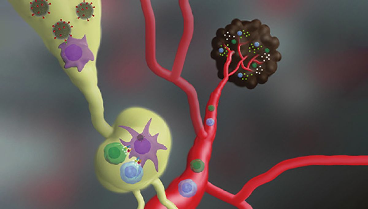
Top: Nano-vaccine mechanism of action: following injection, the nano-vaccine internalizes into immune cells, leading to activation of T cells to recognize and attack melanoma.

Bottom: Prof. Carmit Levy (left) and Dr. Tamar Golan.

Credit for both: Prof. Carmit Levy/AFTA


Antibacterial fillings from TAU may combat recurring tooth decay

New material may prevent one of the costliest and most prevalent bacterial diseases in the world

Tooth decay is among the costliest and most widespread bacterial diseases. Virulent bacteria cause the acidification of tooth enamel and dentin, which, in turn, causes secondary tooth decay.

A new study by Tel Aviv University researchers finds potent antibacterial capabilities in novel dental restoratives, or filling materials. According to the research, the resin-based composites, with the addition of antibacterial nano-assemblies, can hinder bacterial growth and viability on dental restorations, the main cause of recurrent cavities, which can eventually lead to root canal treatment and tooth extractions.


Research for the study was led by Dr. Lihi Adler-Abramovich and TAU doctoral student Lee Schnaider in collaboration with Prof. Ehud Gazit, Prof. Rafi Pilo, Prof. Tamar Brosh, Dr. Rachel Sarig and colleagues from TAU’s Maurice and Gabriela Goldschleger School of Dental Medicine and George S. Wise Faculty of Life Sciences. It was published in ACS Applied Materials & Interfaces on May 28.


Can your fillings fight germ?


“Antibiotic resistance is now one of the most pressing healthcare problems facing society, and the development of novel antimicrobial therapeutics and biomedical materials represents an urgent unmet need,” says Dr. Adler-Abramovich. “When bacteria accumulate on the tooth surface, they ultimately dissolve the hard tissues of the teeth. Recurrent cavities — also known as secondary tooth decay — at the margins of dental restorations results from acid production by cavity-causing bacteria that reside in the restoration-tooth interface.”


This disease is a major causative factor for dental restorative material failure and affects an estimated 100 million patients a year, at an estimated cost of over $30 billion.


Historically, amalgam fillings composed of metal alloys were used for dental restorations and had some antibacterial effect. But due to the alloys’ bold color, the potential toxicity of mercury and the lack of adhesion to the tooth, new restorative materials based on composite resins became the preferable choice of treatment. Unfortunately, the lack of an antimicrobial property remained a major drawback to their use.


“We’ve developed an enhanced material that is not only aesthetically pleasing and mechanically rigid but is also intrinsically antibacterial due to the incorporation of antibacterial nano-assemblies,” Schnaider says. “Resin composite fillings that display bacterial inhibitory activity have the potential to substantially hinder the development of this widespread oral disease.”


From nano materials to major breakthroughs


The scientists are the first to discover the potent antibacterial activity of the self-assembling building block Fmoc-pentafluoro-L-phenylalanine, which comprises both functional and structural subparts. Once the researchers established the antibacterial capabilities of this building block, they developed methods for incorporating the nano-assemblies within dental composite restoratives. Finally, they evaluated the antibacterial capabilities of composite restoratives incorporated with nanostructures as well as their biocompatibility, mechanical strength and optical properties.


“This work is a good example of the ways in which biophysical nanoscale characteristics affect the development of an enhanced biomedical material on a much larger scale,” Schnaider says.


“The minimal nature of the antibacterial building block, along with its high purity, low cost, ease of embedment within resin-based materials and biocompatibility, allows for the easy scale-up of this approach toward the development of clinically available enhanced antibacterial resin composite restoratives,” Dr. Adler-Abramovich says.

 
The researchers are now evaluating the antibacterial capabilities of additional minimal self-assembling building blocks and developing methods for their incorporation into various biomedical materials, such as wound dressings and tissue scaffolds.

TAU scientist elected EMBO member

Prof. Judith Berman will now be a member of the The European Molecular Biology Organization which promotes excellence in the life sciences

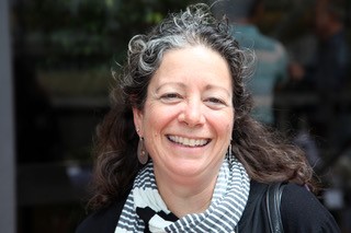
Tel Aviv University’s Prof. Judith Berman was recently named one of 56 new members of the European Molecular Biology Organization (EMBO), joining a group of more than 1800 of the finest researchers in Europe and around the world.
 

Prof. Judith BermanProf. Berman, of TAU’s George S. Wise Faculty of Life Sciences, is being recognized for her outstanding achievements in the study of the growth and evolution of yeast. Prof. Berman, a Fellow of the American Association for the Advancement of Science (AAAS) and the American Society for Microbiology, uses yeasts, especially pathogenic yeasts, to address basic mechanisms of genome change that underlie rapid phenotypic responses to stress.  


“We are so proud of Prof. Berman for being elected to EMBO, which chooses the most exceptional scientists to join their ranks,” says Prof. Karen Avraham, Vice Dean of TAU’s Sackler Faculty of Medicine, a member of the EMBO Council and Prof. Berman’s nominator for EMBO.

“Prof Berman is an appreciated member of the Faculty of Life Sciences at TAU,” says Prof. Abdussalam Azem, Dean of TAU’s Faculty of Life Sciences. “Electing Prof Judith Berman as an EMBO Member is a strong recognition of her research, which is at the forefront of molecular biology of pathogenic yeasts.” 


EMBO Members actively participate in the execution of the organization’s initiatives by sitting on committees and editorial boards, evaluating applications for EMBO funding, mentoring young scientists and providing suggestions and feedback on activities.


“EMBO Members conduct research at the forefront of all life science disciplines, ranging from computational models or analyses of single molecules and cellular mechanics to the study of higher-order systems in development, cognitive neuroscience and evolution,” adds EMBO Director Maria Leptin.


Improving the future of medicine

“It is a great honor to be recognized for my study of pathogenic yeasts of humans and their responses to antifungal drug stress,” says Prof. Berman. “These include mitotic defects that cause aneuploidy and cell-to-cell heterogeneity driven by non-genetic mechanisms.


“We investigate the interplay between chromosome instability, membrane and cell wall dynamics, and intracellular localization of antifungal drugs to better understand processes that modulate the amplitude and diversity of phenotypic responses.”


EMBO will formally welcome its new members and associate members at the Annual Members’ Meeting in Heidelberg between October 29-31.


It’s Simple Math

TAU is pursuing creative ways to get more girls interested in STEM subjects

Whoops and cheers rip through the room as industrial engineering student Merav David looks on with amusement. She has just told 60 teenage girls on a tour of TAU’s Iby and Aladar Fleischman Faculty of Engineering that women are by far the highest achievers among the students in her third-year class. The girls on the tour study advanced math and science in Bat Yam high schools – defying the gender gap in science, technology, engineering and mathematics (STEM). This gap is evident from an early age and carries through university, where women represent fewer than 25% of STEM graduates in developed countries worldwide, even as they outnumber men in study programs overall. Starting young is critical according to Prof. Rachel Gali Cinamon, Head of TAU’s Jaime and Joan Constantiner School of Education: “The current system misses out on girls. We must engage them before they are tracked into non-STEM fields.”


A new TAU program, “Girls Think Science,” is designed to spark girls’ interest in STEM subjects during the golden window of learning from 3rd to 6th grade. Girls from Israel’s social and geographic periphery, including Arabs and Orthodox Jews, enjoy experiential learning at STEM labs, guided by female students and researchers. The program expands upon engineering tours that Dr. Dana Ashkenazi of the School of Mechanical Engineering has been organizing on a volunteer basis for the past decade at TAU’s Engineering Faculty. Girls attending the tours get answers to questions such as “why is the sky blue?” at Prof. Avishay Eyal’s Optics & Photonics Lab, guided by doctoral student Lihi Shiloh; see the inner workings of the body with 3D printing of biological organs at Dr. Orna Sharabani-Yosef’s Tissue Engineering Lab; and encounter artificial intelligence (AI) robots at Dr. Goren Gordon’s Curiosity Lab.


During her PhD studies in the 1990s, Dr. Ashkenazi was the lone woman in a class of 40. This experience motivated her to introduce girls to the joys of science in the hope of recruiting more women to STEM. “My parents exposed me to scientific and engineering topics from a young age. But still, as a mother, I could see my daughters being steered toward humanities tracks at school. I tell them they can do whatever they set their minds to, but society says something else. Girls begin to question themselves, their abilities and their chances to succeed.”


Questioning one’s ability to succeed influences the high rate of attrition that increases with each higher education milestone among women in STEM. Prof. Cinamon, in conjunction with Israel’s Ministry of Science & Technology, studied this phenomenon and developed unique interventions for all stages of academic development, from BSc through post-doc. Interventions range from mentorship to reframing the post-doc as a unique family experience. “Among undergrads, MScs, and PhDs we found that academic identity – rather than academic achievement – is the major factor deter-mining whether a student will pursue higher level STEM studies. Women may have phenomenal grades and academic achievements, yet still may believe they are not good enough.”


The post-doc hurdle

But what are women’s chances of making it in STEM? The TAU President’s Advisor on Gender Equity Prof. Ilana Eli runs the numbers: “Women represent 54% of PhD candidates at TAU, but less than 50% of TAU lecturers and only 22% of professors. In STEM fields these percentages drop precipitously, with some departments employing only one female faculty member among nearly 50 men.


“These numbers reflect the past – professors today began their careers more than two decades ago. Yet these numbers also influence the future – our female students lack role models showing the academic path as accessible to women, especially in STEM,” says Eli. The postdoc is the most formidable obstacle for Israeli women in academia. By the time they complete their PhDs many are starting a family and a postdoc placement of two to four years abroad can seem untenable. TAU is now helping by awarding yearly stipends of $25,000 each for women postdocs in STEM.


“We grant five stipends annually. But deserving candidates are double that number and we wish we could grant more,” says Prof. Eli.


Other programs include a joint post doc in which women conduct research abroad under the auspices of a foreign university as well as at TAU, thus cutting the need for a multiyear relocation. The President’s office also sponsors stipends for travel with a nursing baby and caregiver, enabling new mothers to participate in international conferences essential for establishing themselves in academia.


Changing reality

TAU scientists are eager to boost the number of female faculty through Girls Think Science. “It’s simple math: The larger the pool of girls exposed to STEM, the greater the chance of women choosing to go into STEM fields, both in industry and academia,” says Prof. Shiri Artstein-Avidan, the only female full professor of pure math among over 40 professors at the Raymond and Beverly Sackler School of Mathematical Sciences and a 2016 Kadar Family Award winner. “My father is a mathematician. I want to bring girls who were not brought up in a scientific milieu to this fascinating world.”


Dr. Vered Padler-Karavani of the George S. Wise Faculty of Life Sciences did not enjoy access to science in her home or local school. She grew up in a development town and discovered science through TAU’s long-running Dov Lautman Unit for Science Oriented Youth (now Youth University). “Starting early is important, as is having someone to look up to,” says Padler-Karavani. That is why she volunteers as Chair of ISEF—the Israeli Scholarship Education Foundation—which seeks to expand STEM among youth in Israel’s periphery. She is eager to host Girls Think Science participants in her lab where she studies how to target diseased cells through their protein and sugar coatings.


Role models are crucial, maintains Dr. Ashkenazi. “We hold our tours in the presence of female faculty members and students as mentors.” Ashkenazi believes that the program must engage students from Israel’s periphery, not just from the urban, affluent areas of central Israel because, “When it comes to these subjects, just being a girl places you in the periphery.”


Featured image: High school girls from Bat Yam touring the teaching laboratory at the Department of Materials Science and Engineering at the Iby and Aladar Fleischman Faculty of Engineering
 

New system for detecting Parkinson’s early

New method tracks early stages of protein aggregation involved in Parkinson’s

Parkinson’s disease is a debilitating neurodegenerative disease, affecting everything from speech, posture and gait to digestion, sleep, impulse control and cognition. Therapies exist that alleviate some symptoms of the disease, but there is still no cure for Parkinson’s, which affects close to one million Americans and 10 million people worldwide.

A new Tel Aviv University study unveils a novel method for detecting the aggregation of the protein alpha-synuclein, a hallmark of Parkinson’s disease. With this knowledge, caregivers could introduce treatment that has the potential to significantly delay disease progression.

By the time a patient is diagnosed with Parkinson’s disease, 50 percent to 80 percent of the dopaminergic cells in the part of the brain called substania nigra are already dead, possibly due to development of toxicity as result of alpha-synuclein aggregation. “We have developed a new method for tracking early stages of aggregation of alpha-synuclein using super-resolution microscopy and advanced analysis,” says Prof. Uri Ashery, co-author of the study and head of TAU’s Sagol School of Neuroscience and TAU’s George S. Wise Faculty of Life Sciences. The research was published in Acta Neuropathologica on May 31.

“Together with our collaborators at Cambridge University, who developed a special mouse model for Parkinson’s disease, we were able to detect different stages of the aggregation of this protein,” Prof. Ashery explains. “We correlated the aggregation with the deteriorating loss of neuronal activity and deficits in the behavior of the mice.”

A big step towards early detection

“This is extremely important because we can now detect early stages of alpha-synuclein aggregation and monitor the effects of drugs on this aggregation,” says Dr. Dana Bar-On of the Sagol School of Neuroscience, a co-author of the study. “We hope that this research can be implemented for use in the early diagnosis of Parkinson’s in patients. We’re currently working to implement the methods in a minimally invasive manner with Parkinson’s patients.”

The researchers, in collaboration with the Max Planck Institute in Gottingen and Ludwig-Maximilians-Universität München, were able to illustrate the effect of a specific drug, anle138b, on this protein aggregation and correlated these results with the normalization of the Parkinson’s phenotype in the mice, according to Prof. Ashery. “This is a significant step forward in the world of Parkinson’s research,” he says.

The researchers are planning to expand their research to family members of Parkinson’s disease patients. “By detecting aggregates using minimally invasive methods in relatives of Parkinson’s disease patients, we can provide early detection and intervention and the opportunity to track and treat the disease before symptoms are even detected,” Prof. Ashery concludes.

TAU Scientists Print First 3D Heart Using Patient’s Own Cells and Materials

Engineered heart completely matches the immunological, cellular, biochemical and anatomical properties of the patient

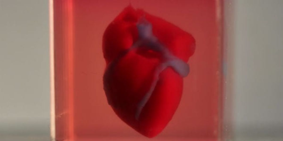
In a major medical breakthrough, Tel Aviv University researchers have “printed” the world’s first 3D vascularised engineered heart using a patient’s own cells and biological materials. Their findings were published on April 15 in a study in Advanced Science.

Until now, scientists in regenerative medicine — a field positioned at the crossroads of biology and technology — have been successful in printing only simple tissues without blood vessels.

“This is the first time anyone anywhere has successfully engineered and printed an entire heart replete with cells, blood vessels, ventricles and chambers,” says Prof. Tal Dvir of TAU’s School of Molecular Cell Biology and Biotechnology, Department of Materials Science and Engineering, Center for Nanoscience and Nanotechnology and Sagol Center for Regenerative Biotechnology, who led the research for the study.

Heart disease is the leading cause of death among both men and women in the United States. Heart transplantation is currently the only treatment available to patients with end-stage heart failure. Given the dire shortage of heart donors, the need to develop new approaches to regenerate the diseased heart is urgent.

“This heart is made from human cells and patient-specific biological materials. In our process these materials serve as the bioinks, substances made of sugars and proteins that can be used for 3D printing of complex tissue models,” Prof. Dvir says. “People have managed to 3D-print the structure of a heart in the past, but not with cells or with blood vessels. Our results demonstrate the potential of our approach for engineering personalized tissue and organ replacement in the future.”

Research for the study was conducted jointly by Prof. Dvir, Dr. Assaf Shapira of TAU’s Faculty of Life Sciences and Nadav Moor, a doctoral student in Prof. Dvir’s lab.


​”At this stage, our 3D heart is small, the size of a rabbit’s heart,” explains Prof. Dvir. “But larger human hearts require the same technology.”

The secret to a new heart

For the research, a biopsy of fatty tissue was taken from patients. The cellular and a-cellular materials of the tissue were then separated. While the cells were reprogrammed to become pluripotent stem cells, the extracellular matrix (ECM), a three-dimensional network of extracellular macromolecules such as collagen and glycoproteins, were processed into a personalized hydrogel that served as the printing “ink.”

After being mixed with the hydrogel, the cells were efficiently differentiated to cardiac or endothelial cells to create patient-specific, immune-compatible cardiac patches with blood vessels and, subsequently, an entire heart.

According to Prof. Dvir, the use of “native” patient-specific materials is crucial to successfully engineering tissues and organs.

“The biocompatibility of engineered materials is crucial to eliminating the risk of implant rejection, which jeopardizes the success of such treatments,” Prof. Dvir says. “Ideally, the biomaterial should possess the same biochemical, mechanical and topographical properties of the patient’s own tissues. Here, we can report a simple approach to 3D-printed thick, vascularized and perfusable cardiac tissues that completely match the immunological, cellular, biochemical and anatomical properties of the patient.”

What organ would you like?

The researchers are now planning on culturing the printed hearts in the lab and “teaching them to behave” like hearts, Prof. Dvir says. They then plan to transplant the 3D-printed heart in animal models.

“We need to develop the printed heart further,” he concludes. “The cells need to form a pumping ability; they can currently contract, but we need them to work together. Our hope is that we will succeed and prove our method’s efficacy and usefulness.


“Maybe, in ten years, there will be organ printers in the finest hospitals around the world, and these procedures will be conducted routinely.”

New genetically encoded sensor isolates hidden Leukemic cells

Cells express surface markers that help them escape most targeted therapies, Tel Aviv University researchers say

Understanding how leukemic stem cells are regulated has become an important area of cancer research. All stem cells can multiply, proliferate and differentiate. Because of these qualities, leukemic stem cells are the most malignant of all leukemic cells. 

A team of Tel Aviv University researchers have now devised a novel biosensor that can isolate and target leukemic stem cells. The research team, led by Dr. Michael Milyavsky of the Department of Pathology at TAU’s Sackler School of Medicine, discuss their unique genetically encoded sensor and its ability to identify, isolate and characterize leukemic stem cells in a study published on January 31 in Leukemia.

Raising the survival rate for blood cancers

“The major reason for the dismal survival rate in blood cancers is the inherent resistance of leukemic stem cells to therapy,” Dr. Milyavsky says. “But only a minor fraction of leukemic cells have high regenerative potential, and it is this regeneration that results in disease relapse. A lack of tools to specifically isolate leukemic stem cells has precluded the comprehensive study and specific targeting of these stem cells until now.”

Until recently, cancer researchers used markers on the surface of the cell to distinguish leukemic stem cells from the bulk of cancer cells, with only limited success. “There are hidden cancer stem cells that express differentiated surface markers despite their stem cell function. This permits those cells to escape targeted therapies,” Dr. Milyavsky explains. “By labeling leukemia cells on the basis of their stem character alone, our sensor manages to overcome surface marker-based issues.

“We believe that our biosensor can provide a prototype for precision oncology efforts to target patient-specific leukemic stem cells to fight this deadly disease.”

Personalized medical testing

The scientists searched genomic databases for “enhancers,” the specific regulatory regions of the genome that are particularly active in stem cells. Then they harnessed genome engineering to develop a sensor composed of a stem cell active enhancer fused with a fluorescence gene that labels the cells in which the enhancer is active.

The scientists were also able to demonstrate that sensor-positive leukemia stem cells are sensitive to a known and inexpensive cancer drug called 4-HPR (fenretinide), providing a novel biomarker for patients who can potentially benefit from this drug.

“Using this sensor, we can perform personalized medicine oriented to drug screens by barcoding a patient’s own leukemia cells to find the best combination of drugs that will be able to target both leukemia in bulk as well as leukemia stem cells inside it,” Dr. Milyavsky concludes. “We’re also interested in developing killer genes that will eradicate specific leukemia stem cells in which our sensor is active.”

The researchers are now investigating those genes that are active in leukemic stem cells in the hope finding druggable targets.

Victoria

Tok Corporate Centre, Level 1,
459 Toorak Road, Toorak VIC 3142
Phone: +61 3 9296 2065
Email: [email protected]

New South Wales

Level 22, Westfield Tower 2, 101 Grafton Street, Bondi Junction NSW 2022
Phone: +61 418 465 556
Email: [email protected]

Western Australia

P O Box 36, Claremont,
WA  6010
Phone: :+61 411 223 550
Email: [email protected]