

TAU Co-Study: “Green Revolution” Decreased Infant Mortality
“Israel, as a global leader in agriculture R&D, has much to offer to the developing world.”
In the first global-scale study of its kind, researchers used wide-scale data to correlate between the “Green Revolution” in agriculture and the dramatic reduction in infant mortality in the developing world. The Green Revolution was a global effort to increase the global crop yield during the second half of the twentieth century.
“In our study, we sought to use empirical methods based on our hypothesis that larger crop larger yields could improve the level of nutrition of pregnant women and young children, and also increase household income, thus contributing indirectly to improved health,” explained Dr. Fishman, of the TAU Department of Public Policy and the Boris Mints Institute for Strategic Policy Solutions to Global Challenges, who contributed to the research. “During the Green Revolution, there was support for international public agricultural R&D with a focus on developing higher-yielding strains of common staple crops, such as wheat, rice, and corn. By the end of the 20th century, approximately 60% of the developing world’s agricultural lands were using these varieties.”
At the same time, between 1960 and 2000, there was a dramatic improvement in health in the developing world- the percentage of children who died before the age of one was reduced from 20% to 10%. The cause of this improvement has been long-contested and attributed to various public health improvements but the contribution of individual factors, including the impacts of the Green Revolution, has been poorly quantified until now.
The correlation in the study suggests that the Green Revolution was responsible for a decline of some 2.5- 5% in the rate of infant mortality. This represents between 25% to 50% of the overall reduction of infant mortality during that time period.
To conduct the study, the researchers collected detailed data about the mortality rates of 600,000 infants born in 37 developing countries between 1961 and 2000, and cross-referenced them with information about the diffusion of the improved Green Revolution seeds in the place and year of birth of each of these infants. Using sophisticated statistical methods, they estimated the association between these two variables. The analysis found a statistically significant causal link between the two data sets. In locations where improved varieties diffused earlier – in part because of the types of crops grown, there was also a more rapid decrease in mortality rates.
“Our study proves the historical importance of agricultural R&D for the health of the rural populations in the developing world. We showed that improved crop varieties, which thus improved nutrition and income and reduced hunger, saved the lives of tens of millions of children in the second half of the twentieth century, and have most likely also brought about improved health for tens of millions of other individuals not directly visible in the data,” said Dr. Fishman.
According to Dr. Fishman, these findings highlight the continued need to address public health. “Israel, as a global leader in agriculture R&D, has much to offer to the developing world,” he says.
The study was conducted by an international team of researchers from Tel Aviv University, the Indian School of Business, the World Bank, the University of California San Diego, Michigan State University, and Colorado State University. The paper was published in the Journal of Health Economics.
Featured images: TAU Prof. Ram Fishman and agricultural expert Omar Zaidan explain seedling use to farmers in India. Credit: the Nitzan Lab


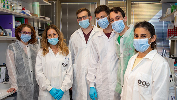


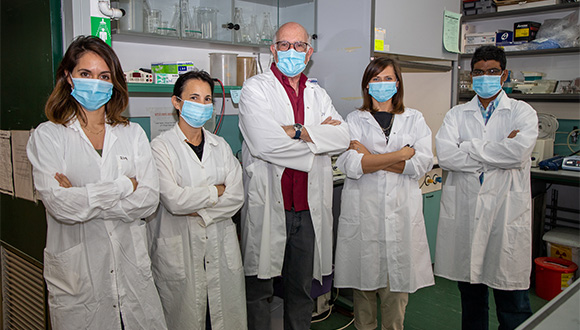 Prof. Gershoni’s Lab team (Photographer: Moshe Bedarshi)
This study is anchored in more than 30 years of research on the interaction of RNA viruses with their receptors and the immune response against them, noted Professor Tal Pupko, Head of the Shmunis School at TAU. “The 3M grant will dramatically accelerate the pace of research for overcoming COVID-19,” said Professor Pupko, adding that Tel Aviv University was particularly proud to be included in this important global initiative by 3M.
“Science is at the heart of 3M and we are committed to advancing the rapid study of this virus as part of our continued effort to combat the COVID-19 pandemic,” said Isabelle Zadikov-Carp, 3M Israel Country Leader. “It’s important that 3M holds true to its core values by supporting our communities and improving lives. We hope that the grant to TAU will facilitate the development of an effective vaccine and we will be keenly following the progress and outcomes of Professor Gershoni’s research with interest.”
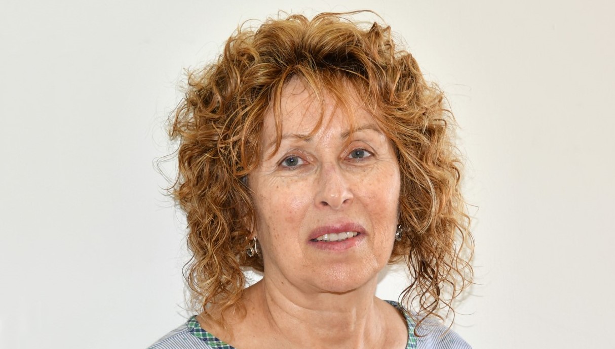
Featured image: Professor Jonathan Gershoni (Photographer: Moshe Bedarshi)
Prof. Gershoni’s Lab team (Photographer: Moshe Bedarshi)
This study is anchored in more than 30 years of research on the interaction of RNA viruses with their receptors and the immune response against them, noted Professor Tal Pupko, Head of the Shmunis School at TAU. “The 3M grant will dramatically accelerate the pace of research for overcoming COVID-19,” said Professor Pupko, adding that Tel Aviv University was particularly proud to be included in this important global initiative by 3M.
“Science is at the heart of 3M and we are committed to advancing the rapid study of this virus as part of our continued effort to combat the COVID-19 pandemic,” said Isabelle Zadikov-Carp, 3M Israel Country Leader. “It’s important that 3M holds true to its core values by supporting our communities and improving lives. We hope that the grant to TAU will facilitate the development of an effective vaccine and we will be keenly following the progress and outcomes of Professor Gershoni’s research with interest.”
Featured image: Professor Jonathan Gershoni (Photographer: Moshe Bedarshi)
