
Tag: Medicine

Is This the Future of Burn Care?

TAU researchers and Sheba Medical Center developed bioengineered skin that accelerates healing time.

Researchers from Tel Aviv University and Sheba Tel Hashomer Medical Center have developed an innovative bioengineered skin equivalent for grafting in burn victims. The bioengineered skin produced from the patient’s cells is more stable, robust, and flexible than current treatments, making it easier to handle. A full-thickness wound model, it achieved wound closure in half the time of standard therapies. This pioneering study was driven by the ongoing war and the surge in severe burn injuries, which underscored the urgent need for better treatment solutions.

The groundbreaking study was led by Prof. Lihi Adler-Abramovich and PhD student Dana Cohen-Gerassi from the Laboratory for Bio-Inspired Materials and Nanotechnology at the Goldschleger School of Dental Medicine, at TAU’s Gray Faculty of Medical and Health Sciences, in collaboration with: Dr. Ayelet Di Segni, Director of the Sheba Tissue Bank and the Green Skin Engineering Laboratory at Sheba; Dr. Amit Sitt from TAU’s School of Chemistry, Faculty of Exact Sciences, Prof. Josef Haik, Head of the Plastic Surgery Division and National Burn Center at Sheba; Dr. Moti Harats, Head of the Israeli National Intensive Care Burn Center at Sheba; Dr. Marina Ben-Shoshan and Dr. Adi Liiani scientists from the Green Skin Engineering Laboratory at Sheba; Prof. Itzhak Binderman from TAU’s Goldschleger School of Dental Medicine and Prof. Yosi Shacham-Diamand from TAU’s Fleischman Faculty of Engineering, as well as PhD candidate  Tomer Reuveni and Offir Loboda from TAU. The paper was published in the prestigious scientific journal Advanced Functional Materials.

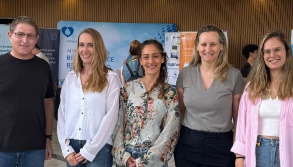
The research team from Tel Aviv University and Sheba Medical Center (From left to right): Dr. Amit Sitt, Dr. Marina Ben-Shoshan, Dr. Ayelet Di Segni, Prof. Lihi Adler-Abramovich & Dana Cohen-Gerassi.

“Surgical intervention is often essential for second-degree burns and above to restore skin, prevent infection, and save lives,” explains Prof. Lihi Adler-Abramovich. “The current gold-standard treatment is ‘autologous skin grafting’, in which healthy skin is harvested from another area of the patient’s body and transplanted onto the burn site. However, this approach has significant disadvantages, particularly the need to damage healthy tissue to treat the injury. This becomes especially problematic in cases of extensive burns, where the availability of intact skin is limited.”

Smart Skin That Heals You Faster

“One of the most advanced alternatives, currently offered in Israel only at Sheba Medical Center, is ‘cultured epidermal autograft’ (CEA). Instead of removing a large skin section, a small biopsy is taken, and cells extracted from that sample are cultured in the lab to produce skin grafts for transplantation. While this method avoids donor-site damage, it comes with several challenges: First, the skin cells are grown on a layer of mouse-derived feeder cells, requiring strict regulation to ensure no mouse cells remain in the graft; Second, once removed from the culture dish, the CEA shrinks by over 50%, significantly reducing yield up to 30 grafts may be needed to cover a single area, such as an arm or leg. Finally, the lab-grown skin consists of only the upper epidermal layer, making it extremely thin, fragile, and prone to curling at the edges.”

The need for advanced solutions is particularly urgent in wartime, with many soldiers suffering from burns. For both soldiers and civilians, a durable bioengineered graft could significantly improve chances for recovery and a good quality of life. “Since October 2023, Sheba has treated many young people with burn injuries,” says Dr. Ayelet Di Segni. “At such a time, bringing knowledge accumulated in the lab directly to the patient’s bedside becomes an urgent and tangible goal. We aim to develop a graft that can truly transform the process of recovery.”

Made from Your Own Cells

To address this challenge, researchers from Tel Aviv University and Sheba Medical Center collaborated to develop multi-cellular, multi-layered bioengineered skin grafts designed to mimic the properties and function of natural skin, without shrinking,  tearing upon contact, or relying on animal-derived additives.

“We designed a nanofiber scaffold made of a polymer called PCL, which is already FDA-approved, and combined it with a bioactive peptide – a short amino acid sequence that promotes cell adhesion, growth, and proliferation,” explains PhD student Dana Cohen-Gerassi. “We then seeded this scaffold with skin cells derived from a patient’s biopsy. Remarkably, the cells organized themselves naturally: fibroblasts populated one side of the scaffold, while keratinocytes grew on the other – mimicking the structure of real human skin.”

Dr. Marina Ben-Shoshan, senior researcher at Sheba’s Green Center for Skin Graft Engineering, adds: “Our graft is unique in that it does not shrink, and is durable, flexible, and easy to handle. Implantation in model animals has yielded impressive results, accelerating the healing process. While the standard treatment closes half of the burn wound in eight days, with our method, this took only four days. Moreover, we observed that essential skin structures, such as hair follicles, began to grow.”

Dr. Amit Sitt from TAU’s School of Chemistry adds: “The nanofiber scaffolds are made from easily available biocompatible materials and produced via a scalable spinning process. In the future, this will enable large-scale production of fiber sheets, as well as incorporation of additional substances to facilitate the healing process.”

Prof. Yossi Haik of Sheba Medical Center concludes: “The bioengineered skin we’ve developed represents a true breakthrough in burn care. Made entirely from the patient’s cells, it is strong, flexible, easy to handle, and significantly accelerates healing. This is a major step towards personalized therapies that can greatly improve the recovery and quality of life of severe burn victims, both soldiers and civilians. In the next phase, we plan to conduct trials in additional models and advance the necessary regulatory processes to bring this innovative technology closer to clinical application.”

Can AI Help Doctors Make Better Diagnoses?

A new TAU study explores how accurate AI can be when assisting with diagnoses in virtual urgent care.

A new study led by Prof. Dan Zeltzer, a digital health expert from the Berglas School of Economics at Tel Aviv University, compared the quality of diagnostic and treatment recommendations made by artificial intelligence (AI) and physicians at Cedars-Sinai Connect, a virtual urgent care clinic in Los Angeles, operated in collaboration with Israeli startup K Health. The paper was published in Annals of Internal Medicine and presented at the annual conference of the American College of Physicians (ACP). This work was supported by funding from K Health.

AI vs. Physicians in Virtual Care

Prof. Zeltzer explains: “Cedars-Sinai operates a virtual urgent care clinic offering telemedical consultations with physicians specializing in family and emergency care. Recently, an AI system was integrated into the clinic—an algorithm based on machine learning that conducts initial intake through a dedicated chat incorporates data from the patient’s medical record and provides the attending physician with detailed diagnostic and treatment suggestions at the start of the visit -including prescriptions, tests, and referrals. After interacting with the algorithm, patients proceed to a video visit with a physician who ultimately determines the diagnosis and treatment. To ensure reliable AI recommendations, the algorithm—trained on medical records from millions of cases—only offers suggestions when its confidence level is high, not recommending about one out of five cases. In this study, we compared the quality of the AI system’s recommendations with the physicians’ actual decisions in the clinic”.


Prof. Dan Zeltzer (Photo courtesy of Richard Haldis).

The researchers examined a sample of 461 online clinic visits over one month during the summer of 2024. The study focused on adult patients with relatively common symptoms—respiratory, urinary, eye, vaginal and dental. In all visits reviewed, patients were initially assessed by the algorithm, which provided recommendations, and then treated by a physician in a video consultation. Afterward, all recommendations—from both the algorithm and the physicians—were evaluated by a panel of four doctors with at least ten years of clinical experience, who rated each recommendation on a four-point scale: optimal, reasonable, inadequate, or potentially harmful. The evaluators assessed the recommendations based on the patient’s medical history, the information collected during the visit, and transcripts of the video consultations.

AI Proves More Accurate Than Physicians in Study

The compiled ratings led to interesting conclusions: AI recommendations were rated as optimal in 77% of cases, compared to only 67% of the physicians’ decisions; at the other end of the scale, AI recommendations were rated as potentially harmful in a smaller portion of cases than physicians’ decisions (2.8% of AI recommendations versus 4.6% of physicians’ decisions).  In 68% of the cases, the AI and the physician received the same score; in 21% of cases, the algorithm scored higher than the physician; and in 11% of cases, the physician’s decision was considered better.

The explanations provided by the evaluators for the differences in ratings highlight several advantages of the AI system over human physicians: First, the AI more strictly adheres to medical association guidelines—for example, not prescribing antibiotics for a viral infection; second, AI more comprehensively identifies relevant information in the medical record—such as recurrent cases of a similar infection that may influence the appropriate course of treatment; and third, AI more precisely identifies symptoms that could indicate a more serious condition, such as eye pain reported by a contact lens wearer, which could signal an infection. Physicians, on the other hand, are more flexible than the algorithm and have an advantage in assessing the patient’s actual condition. For example, if a COVID-19 patient reports shortness of breath, a doctor may recognize it as relatively mild respiratory congestion, whereas the AI, based solely on the patient’s answers, might refer them unnecessarily to the emergency room.

A Step Closer to Supporting Doctors

Prof. Zeltzer concludes: “In this study, we found that AI, based on a targeted intake process, can provide diagnostic and treatment recommendations that are, in many cases, more accurate than those made by physicians. One limitation of the study is that we do not know which physicians reviewed the AI’s recommendations in the available chart, or to what extent they relied on the recommendations. Thus, the study only measured the accuracy of the algorithm’s recommendations and not their impact on the physicians. The study’s uniqueness lies in the fact that it tested the algorithm in a real-world setting with actual cases, while most studies focus on examples from certification exams or textbooks. The relatively common conditions included in our study represent about two-thirds of the clinic’s case volume, thus the findings can be meaningful for assessing AI’s readiness to serve as a decision-support tool in medical practice. We can envision a near future in which algorithms assist in an increasing portion of medical decisions, bringing certain data to the doctor’s attention, and facilitating faster decisions with fewer human errors. Of course, many questions remain about the best way to implement AI in the diagnostic and treatment process, as well as the optimal integration between human expertise and artificial intelligence in medicine”.

Other authors involved in the study include Zehavi Kugler, MD; Lior Hayat, MD; Tamar Brufman, MD; Ran Ilan Ber, PhD; Keren Leibovich, PhD; Tom Beer, MSc; and Ilan Frank, MSc. Caroline Goldzweig, MD MSHS, and Joshua Pevnick, MD, MSHS.

Why We Really Exercise: First AI Study Reveals True Motivation

A first-of-its-kind AI study finds 23.9% exercise for appearance and 18.9% for health.

A new study from Tel Aviv University used AI tools for the first time to discover what motivates people to exercise and which strategies are most effective for maintaining physical fitness.

The researchers used tools of artificial intelligence and machine learning to scan thousands of posts on the Reddit social network. They found that 23.9% of the users who engage in sports do so to improve their appearance, 18.9% exercise to maintain their physical health, and 16.9% exercise to maintain their mental health.

The study was led by a team of researchers from TAU’s School of Public Health, Faculty of Medical and Health Sciences : Dr. Michal Shmueli-Scheuer, Yedidya Silverman, Prof. Israel Halperin, and Prof. Yftach Gepner. The paper was published in the Journal of Medical Internet Research (JMIR).

Why Don’t We Exercise More, Even When We Know It’s Crucial?

Prof. Gepner explains: “Researchers in our field usually rely on cumbersome old-school questionnaires, containing inherent biases, to understand why people engage in sports and what strategies help them adhere to physical activity.  It’s an astonishing phenomenon: science tells us that if we put just over two hours a week into physical activity, we can prevent 30% of diseases, improve our quality of life, and extend our lifespan; and yet, less than a quarter of the population actually does this. Why? What have we failed to see?  While we all wish our loved ones good health on their birthday, a wish for ‘good workouts’ is quite rare… But there is a way to be healthy – by exercising. That’s why it’s crucial to understand what really motivates people to engage in physical activity and what helps them stick with it”.

“Our findings are not based on self-reporting, a representative sample, a questionnaire, or a survey. This is, in plain terms, the real reason why people exercise. And the answer is that people mainly exercise to look good. In questionnaires, people claim they want to be healthy, but in reality, they want six-pack abs. These findings are important because they teach us how to address the public, how to persuade people to get off the couch, promote health, and prevent disease”, he adds.

Beyond the question of motivation, the researchers also sought to identify strategies that induce people to engage in physical activity. According to the Reddit posts, 30% rely on workout habits (e.g. morning/evening, every Saturday morning), 13.9% set goals (such as losing weight or running 5 km), 12.1% enjoy the activity itself, 9.7% enjoy socializing during workouts, 8.9% use media (such as YouTube workout videos), 2.8% use fitness apps, and 2.5% have made a financial commitment to adhere to physical activity.

“The results are quite significant”, explains Prof. Gepner. “One strategy is more successful and therefore more recommended than others—creating exercise habits. If you want to be healthier, you need to develop healthy habits, period. Instead of a morning cigarette, drink two glasses of water and go out for a run. 30% is an empirical statistic that is hard to argue with, so as the Head of the Department of Health Promotion, I can confidently say to the public: develop habits and be healthy”.

What Happens Inside a Cell Under Stress?

A novel AI-based method reveals how cells respond to drug treatments.

Researchers from Tel Aviv University have developed an innovative method that can help to understand better how cells behave in changing biological environments, such as those found within a cancerous tumor. The new system, called scNET, combines information on gene expression at the single-cell level with information on gene interactions, enabling the identification of important biological patterns such as responses to drug treatments. The scientific article published in the Nature Methods journal explains how scNET may improve medical research and assist in the development of treatments for diseases. The research was led by PhD student Ron Sheinin under Prof. Asaf Madi, from the Faculty of Medical and Health Sciences and Prof. Roded Sharan, head of the School of Computer Science and AI at Tel Aviv University.

From noisy data to clear insights

Today, advanced sequencing technologies allow the measurement of gene expression at the single-cell level and, for the first time, researchers can investigate the gene expression profiles of different cell populations within a biological sample and discover their effects on the functional behavior of each cell type. One fascinating example is understanding the impact of cancer treatments – not only on the cancer cells themselves but also on the pro-cancer supporting cells or anti-cancer cell populations, such as some cells of the immune system surrounding the tumor.

Despite the amazing resolution, these measurements are characterized by high levels of noise, which makes it difficult to identify precise changes in genetic programs that underlie vital cellular functions. This is where scNET comes into play.

A social network for genes

Ron Sheinin: “scNET integrates single-cell sequencing data with networks that describe possible gene interactions, much like a social network, providing a map of how different genes might influence and interact with each other. scNET enables more accurate identification of existing cell populations in the sample. Thus, it is possible to investigate the common behavior of genes under different conditions and to expose the complex mechanisms that characterize the healthy state or response to treatments”.

Prof. Asaf Madi: “In this research, we focused on a population of T cells, immune cells known for their power to fight cancerous tumors. scNET revealed the effects of treatments on these T cells and how they became more active in their cytotoxic activity against the tumor, something that was not possible to discover before due to the high level of noise in the original data”.

Prof. Roded Sharan: “This is an excellent example of how artificial intelligence tools can help decipher biological and medical data, allowing us to gain new and significant insights. The idea is to provide biomedical researchers with computational tools that will aid in understanding how the body’s cells function, thereby identifying new ways to improve our health”.

In conclusion, scNET demonstrates how combining AI with biomedical research could lead to the development of new therapeutic approaches, reveal hidden mechanisms in diseases, and propose new treatment options.

Can Stem Cells Restore Bone Marrow?

Doubling adult stem cells for bone marrow and immune system regeneration.

An international research team, led by scientists from Tel Aviv University and Sheba Medical Center, has unveiled an innovative method for activating adult stem cells from human bone marrow, enabling their expansion outside the body for use in bone marrow regeneration and the construction of a new blood and immune system.

The findings, published in the prestigious journal Nature Immunology, represent a breakthrough that could significantly improve transplant success rates for patients who have undergone intensive chemotherapy, suffer from genetic disorders, or require a bone marrow transplant but are unable to source a sufficient number of stem cells from a donor.

The study was led by Dr. Tomer Itkin from the Faculty of Medical and Health Sciences and the Sagol Center for Regenerative Medicine at Tel Aviv University, and the Neufeld Cardiac Research Institute at Sheba Medical Center, Tel Hashomer. The research also included contributions from leading medical institutions worldwide, including Weill Cornell Medical College and Hospital in New York, the Memorial Sloan Kettering Cancer Center (MSKCC), Mount Sinai Hospital, the University of Toronto Medical Center, and the Fred Hutchinson Cancer Research Center in Seattle.

Dr. Tomer Itkin.

Switching On Stem Cells

In the study, which is based on a comprehensive big data analysis of RNA sequencing and epigenetic DNA sequencing, the researchers identified a key protein—the Fli-1 transcription factor—that activates stem cells of the immune and blood system. These stem cells are highly active when sourced from umbilical cord blood but remain in a “dormant” and inactive state when obtained from adult bone marrow donors. Using modified mRNA technology—the same technology used to develop COVID-19 vaccines—the researchers successfully “awakened” the adult stem cells, allowing them to divide in a controlled manner without cancer risk. The activated cells were expanded on endothelial cells, which mimic the blood vessels that support stem cells in the bone marrow environment, demonstrating an enhanced ability to integrate and restore blood production under transplant conditions.

According to Dr. Itkin, This new method significantly expands the available pool of stem cells for transplantation without relying on rare bone marrow donors. Additionally, the method can be used to treat patients whose stem cells have undergone genetic correction, such as those with thalassemia and hereditary anemia, as well as patients who have undergone multiple rounds of chemotherapy and have an insufficient number of stem cells for autologous transplantation“.

The key takeaway from the study is that activating stem cells through molecular programming, rather than arbitrary cell transplantation, substantially improves the success rates of regenerative treatments. The next stage of research involves testing the method in clinical trials to bring this groundbreaking technology into widespread therapeutic use. Furthermore, the researchers plan to apply the same therapeutic approach to regenerate additional tissues, including those without existing adult stem cells, such as the heart.


New PTEN Research Could Transform Autism and Cancer Studies

Is PTEN the key to advancing autism and cancer research?

A novel scientific method developed at Tel Aviv University promises to accelerate our understanding of the gene PTEN, a key player in cellular growth. This breakthrough will help scientists better understand how cells grow and divide, potentially leading to advancements in the treatment of various conditions, including developmental disorders and various forms of cancer.

The study, led by Dr. Tal Laviv in the Faculty of Medical and Health Sciences at Tel Aviv University, was published in the prestigious journal Nature Methods.

The research team explains that cells in the human body constantly adjust their size and rate of division to adapt to their environment throughout life. This process is crucial for normal development, as cells go through periods of precise growth regulation. When this process is disrupted, it can lead to severe diseases such as cancer and developmental disorders.

In the brain, regulating cellular growth is especially critical during early brain development, which occurs in the first years of life. Many genes are involved in this regulation, but one gene in particular—PTEN (Phosphatase and Tensin Homologue)—plays a central role. Mutations in PTEN are linked to a variety of conditions, including autism, epilepsy, and cancer.

PTEN’s Impact Explained

Dr. Tal Laviv explains: “Many studies have shown that PTEN is essential for regulating cell growth in the brain by providing a stop signal. This means PTEN activity is crucial for maintaining cells at their proper size and state. There is growing evidence that mutations in PTEN, which reduce its activity, contribute to diseases like autism, macrocephaly, cancer, and epilepsy. Despite the critical role PTEN plays in cellular function, scientists have had limited tools to measure its activity. For example, it wasn’t to directly measure PTEN activity in an intact brain, which would greatly help our understanding of its role in health and disease”.

Dr. Laviv and his research team, led by MD-PhD student Tomer Kagan, have developed an innovative tool that directly measures PTEN activity with high sensitivity in various research models, including in the intact brains of mice. This groundbreaking technology, which combines advancements in genetic tools and microscopy, will allow scientists to gain deeper insights into why PTEN is so crucial for normal brain development. It could also improve our understanding of how PTEN-related diseases, such as cancer and autism, develop.

The researchers predict that this new tool will enable the development of personalized therapeutics by monitoring PTEN activity in various biological settings. Additionally, it could help identify diseases at earlier stages, potentially leading to faster and more effective treatments.

Gender-Specific Damage in Rare Genetic Syndrome

.ADNP Protein Causes Different Brain Damage in Males and Females.

Researchers at Tel Aviv University, led by Prof. Illana Gozes, examined the effects of different mutations in the ADNP protein, which is essential for normal brain development and aging, on the brain cells of mice — distinguishing between males and females. To their surprise, they found that the defective protein affects completely different mechanisms in the two sexes: in males, the damage occurs in a mechanism that protects the structure of proteins, which in turn disrupts the process of neurogenesis — the production of new brain cells from stem cells — a process crucial for memory and learning. In females, on the other hand, the mechanism that regulates energy within the cell is impaired, preventing the brain from receiving sufficient energy. All of these processes are essential for maintaining memory and learning functions, and their disruption causes significant impairment in both sexes, leading to the development of incurable brain diseases such as Alzheimer’s, in which ADNP is also found to be defective.

The research was conducted by Prof. Illana Gozes, Dr. Gidon Karmon, and doctoral student Guy Shapira from the and the  at Tel Aviv University. Additional contributors to the study include Prof. Noam Shomron, Dr. Gal Hacohen-Kleiman, doctoral student Maram Ganaiem from the Faculty of Medical and Health Sciences, Dr. Shula Shazman from the Department of Mathematics and Computer Science at the Open University, and researchers from the University Hospital of Thessaloniki in Greece. The study was published in the prestigious journal Molecular Psychiatry from Nature.

Prof. Illana Gozes.

Prof. Gozes stated: “The ADNP protein was discovered in my lab, and we have been researching it for many years. We found that it is critical for brain development and plays a protective role in neurodegenerative diseases like Alzheimer’s. Additionally, it was found that defects in the ADNP gene cause ADNP syndrome, a rare genetic disorder associated with developmental delays, learning disabilities, and symptoms of autism. In parallel, we are developing the experimental drug Davunetide, which is based on a fragment of the ADNP protein. In this study, we aimed to examine whether ADNP is involved in the process known as ‘neurogenesis’ — the formation of new neurons from stem cells in the adult brain, a process essential for memory and learning. We focused on the hippocampus, a brain region crucial for memory, in adult mice”.

Using genetic engineering, the researchers established two mouse models reflecting different forms of ADNP syndrome: mice that express only half the normal amount of ADNP, with only one active allele in the DNA instead of two, which are typically inherited from both parents and mice with a mutation in the ADNP gene that truncates the protein production process, resulting in a shorter-than-normal ADNP protein.

The researchers note that the most severely affected children with ADNP syndrome are those with the mutation that produces the truncated protein. Additionally, neurogenesis was examined in a control group of healthy mice.

To track the course of neurogenesis, a substance was injected into the mice, staining the DNA of brain cells participating in the process. The data were analyzed using computational bioinformatics methods, proving that ADNP plays a crucial role in neurogenesis. Furthermore, a significant difference was found between how ADNP functions in males versus females. First, in healthy mice, neurogenesis was more active in males than females, while in male mice with an ADNP mutation, neurogenesis was reduced to the same level as in females. A fundamental difference between the sexes was also identified in an additional research method: RNA sequencing of all genes in the hippocampus of mice with the truncated ADNP protein.

How ADNP Protein Breaks Brains by Gender

Prof. Gozes explains: “There was almost no overlap. The damage to the ADNP protein affected completely different mechanisms in male and female brains. The explanation for this phenomenon is that in males, one of the functions of ADNP is to regulate a mechanism that maintains protein structure (unfolded protein response), which in turn regulates neurogenesis. The ADNP gene is a master regulator of this entire mechanism in male brains, and when it is defective, the process is significantly impaired. In females, however, the ADNP protein enters the mitochondria — the cell’s energy powerhouse — and when the mutation alters the protein’s structure, less ADNP can enter the mitochondria. As a result, energy production in the cell is likely impaired, disrupting brain function, which requires a large amount of energy”.

As part of the study, the researchers also tested the effectiveness of the experimental drug Davunetide, based on the NAP fragment of the ADNP protein, in treating affected mice. They observed a positive effect in all cases, with particularly significant neurogenesis recovery in the model where mice had only half the normal ADNP levels.

Promising Drug for ADNP and Beyond

Prof. Gozes concludes: “Our research shows that ADNP is closely linked to neurogenesis and that it functions differently in males and females — a finding that has also emerged in previous studies. Additionally, we found that Davunetide, the drug that we discovered and are developing, is effective. We aim to soon begin a clinical trial in children with ADNP syndrome (ADNP deficiency). We hope that in the future, the drug will also help Alzheimer’s patients — in whom we previously found sex-based differences — as well as other neurodegenerative diseases where ADNP is impaired. Notably, the rare and incurable disease Progressive Supranuclear Palsy (PSP), which has pathological similarities to Alzheimer’s disease, showed significant improvement in women treated with Davunetide in our previous clinical study”.

The pharmaceutical development is being carried out by ExoNavis Therapeutics Ltd under a licensing agreement with Ramot, Tel Aviv University’s technology transfer company. Prof. Gozes serves as Vice President for Drug Development at the company.

Half the Tumors Gone: TAU Scientists Remove a Cancer Gene with CRISPR

This gene-editing success from TAU could change cancer treatment forever.

Researchers from Tel Aviv University utilized CRISPR to cut a single gene from cancer cells of head and neck tumors – and successfully eliminated 50% of the tumors in model animals. This groundbreaking study was led by Dr. Razan Masarwy, MD, Ph.D. from the lab of  Prof. Dan Peer – a global pioneer in mRNA-based drugs, Director of the Laboratory of Precision Nanomedicine, VP for Research and Development and member of the Shmunis School of Biomedicine and Cancer Research – all at TAU. The findings were published in the prestigious journal Advanced Science.

A New Approach to Treating Head and Neck Cancers

“Head and neck cancers are prevalent, ranking fifth in cancer mortality”, says Prof. Peer. “These are localized cancers, typically starting in the tongue, throat, or neck, which can later metastasize. If detected early, localized treatment can effectively target the tumor. We aimed to use genetic editing of a single gene expressed in this type of cancer to collapse the entire pyramid of the cancerous cell. This gene is the cancer-specific SOX2, also expressed in other types of cancer and overexpressed in these particular tumors”.

Prof. Dan Peer.

Prof. Peer and his colleagues are global pioneers in developing mRNA-based drugs encased in synthetic lipid particles that mimic biological membranes. In this study, the researchers synthesized special lipids that encapsulate the delivered CRISPR system in an RNA format. An antibody targeting a receptor against a protein named EGF was attached to the surface of these particles.

“These tumors are highly targeted”, explains Prof. Peer. “We targeted EGF because the cancer cells express the EGF receptor. Using our nano-lipid delivery system, we injected the drug directly into the tumor in a tumor model and successfully took out the gene – cutting it out from the cancer cell’s DNA with the CRISPR ‘scissors’. We were happy to observe the domino effect we had predicted. Following three injections spaced one week apart, 50% of the cancerous tumors simply disappeared after 84 days – which did not happen in the control group”.

Prof. Dan Peer & research team.

TAU Pioneers CRISPR for Cancer Treatment

In 2020, Prof. Peer and his team were the first in the world to use CRISPR to cut genes from cancer cells in mice and a cell-specific manner, and this is the first time they have applied it to head and neck cancers.

“Generally, CRISPR isn’t used for cancer because the assumption is that knocking out one gene wouldn’t collapse the whole pyramid. In this study we demonstrated that there are some genes without which a cancer cell cannot survive, making them excellent targets for CRISPR therapy. Since cancer cells sometimes compensate with other genes, it’s possible that additional genes need to be cut out, or perhaps not. Theoretically, this approach could be effective against many types of cancer cells, and we are already working on additional cancer types, including myeloma, lymphoma, and liver cancer”.

This study was supported in part by the EXPERT project (European Union’s Horizon 2020 research and innovation programme (under grant agreement # 825828), and the Shmunis Fund for gene editing.

First of Its Kind: mRNA Drugs Delivered Straight to the Gut

Potential new therapy for Crohn’s, colitis, and other inflammatory diseases.

Researchers at Tel Aviv University have achieved a breakthrough in drug delivery: they have successfully transported lipid nanoparticles encapsulating messenger RNA (mRNA) to the immune system of the small and large intestines — bypassing the liver upon systemic administration. By simply altering the composition of the nanoparticles, the researchers demonstrated that mRNA-based drugs can be directed straight to target cells, avoiding the liver.

The groundbreaking Tel Aviv University study was led by post-doctoral fellow Dr. Riccardo Rampado together with Vice President for R&D Prof. Dan Peer, a pioneer in the development of mRNA therapeutics and Director of the Laboratory of Precision Nano-Medicine at the Shmunis School of Biomedicine and Cancer Research. The study was published on the cover of the prestigious journal Advanced Science.

Prof. Dan Peer.

Targeting Drugs More Precisely with Lipid Nanoparticles

“Everything injected into the bloodstream eventually ends up in the liver — that’s just how our anatomy works”, explains Prof. Peer. “This poses two challenges. First, drugs intended to target specific cells in particular organs may be toxic to the liver. Second, we don’t want drugs to get ‘stuck’ in the liver. Ideally, the drug would reach the target organ first, and any remnants would then break down in the liver. We discovered that altering the proportions of lipids comprising the nanoparticles determines their destination in the bloodstream. This is a general phenomenon, meaning it works regardless of the specific lipids, which makes this a significant breakthrough”.

To demonstrate the concept, Prof. Peer and his team encoded the anti-inflammatory protein interleukin-10 into mRNA, encapsulated it in lipid nanoparticles with a composition different from those typically used (such as in mRNA COVID-19 vaccines), and successfully delivered it to the intestines of animal models with Crohn’s disease and colitis via intravenous injection.

“Not only were we able to deliver an mRNA-based anti-inflammatory drug directly to the inflamed intestine and improve all markers of colitis and Crohn’s disease, but we also transformed the immune cells in the intestine into factories for producing the anti-inflammatory interleukin-10”, Prof. Peer explains. “But this is just a proof of concept study. By tweaking the nanoparticle composition, we could deliver other RNA-based drugs to different organs. There’s a saying in American English: ‘It’s all in the formulation’. That’s exactly what this is about”.

Higher Phospholipids, Faster Delivery

In general, lipid-based drugs are encased in synthetic lipid nanoparticles, which mimic biological membranes. One of these lipids is phospholipid named phosphatidylcholine, a component found in all biological membranes. In vaccines like the COVID-19 vaccine, the mRNA is encapsulated in lipid particles containing about 10% of this phospholipid. Prof. Peer and his team increased the phospholipid ratio to 30% and demonstrated that this adjustment caused the particles to float through the bloodstream like oil on water.

“That’s the whole trick”, Prof. Peer concludes. “We adjusted the lipid composition and found that at 30% phospholipid, the drug is directed straight to the intestine. Of course, this wasn’t a blind trial-and-error approach. We understand the mechanism, at least partially, and recognize that this ratio more closely resembles a natural biological membrane, which intestinal cells are better suited to absorb. Now, we are exploring further adjustments to target the pancreas and other organs that can only be reached by fine-tuning the lipid nanoparticle composition. This direct delivery method for mRNA drugs opens up broad possibilities for developing new and more precise therapies than ever before”.

What Happens When the Brain Learns Two Behaviors at Once?

TAU researchers reveal the brain resolves conflict by blocking dual learning.

A new study from Tel Aviv University could reshape our scientific understanding of how humans learn and form memories, particularly through classical and operant conditioning. The research team found that our brain engages in fierce competition between these two learning systems and that only one can prevail at any given time. If we try to learn two conflicting actions for the same situation simultaneously, the result will be confusion, making it difficult to perform either action when re-encountering the situation. In their study, the researchers demonstrated this phenomenon in fruit flies. When the flies were trained to associate a smell with a randomly delivered electric shock (classical conditioning) and also to connect their actions to the smell and shock (operant conditioning), the flies became confused and failed to exhibit a clear response to the shock.

The intriguing research was led by Prof. Moshe Parnas and PhD student Eyal Rozenfeld from the Laboratory for Neural Circuits and Olfactory Perception at Tel Aviv University’s Faculty of Medical and Health Sciences. The findings were published in the prestigious journal Science Advances.

The researchers explain that humans learn in a variety of ways. A well-known example of learning is Ivan Pavlov’s famous experiment, where a dog learns to associate the sound of a bell with food. This type of learning is called classical conditioning and involves passive associations between two stimuli. On the other hand, humans can also learn from their own actions: if a specific action produces a positive outcome, we learn to repeat it, and if it harms us, we learn to avoid it. This type of learning is called operant conditioning and involves active behavior.

Freeze or flee? Cracking the brain’s decision code

For many years, scientists believed that these types of memory work together in the brain. But what happens if the two memories dictate conflicting actions? For instance, mice can be trained to fear a certain smell using both conditioning methods, but their responses will differ depending on which method is employed. Under classical conditioning, the mice will freeze in place, while under operant conditioning, they will flee. What happens if both memories are present simultaneously? Will the mice freeze, flee, or simply continue behaving as if nothing happened?

In a unique study conducted on fruit flies (Drosophila), researchers at Tel Aviv University discovered that the brain cannot learn using both classical and operant conditioning simultaneously. The brain actively suppresses the formation of both types of memories at the same time, using this strategy to determine which behavior to execute. During the experiment, the researchers taught the flies to associate a smell with an electric shock.  When classical conditioning was used flies learned to freeze when they smelled the conditioned odor. In contrast, when operant conditioning was used, flies learned to flee from the smell to avoid the electric shock. They demonstrated that the flies could not learn both lessons together and that attempts to teach both types of learning simultaneously led to no learning at all. Furthermore, they identified the brain mechanisms that prioritize one type of learning over the other.

“Our research completely changes the way we have thought for decades about how our brain learns,” explains Prof. Parnas. “You can think of the brain as engaging in a ‘mental tug-of-war’: if you focus on learning through your actions, the brain blocks the formation of automatic associations. This helps avoid confusion but also means you can’t learn two things simultaneously”.

Why multitasking makes you forget

Prof. Parnas adds: “Fruit flies have simple brains, which makes them easy to study, but their brains are surprisingly similar to those of mammals—and thus to our own. Using powerful genetic tools, the researchers gained a deep understanding of how different learning systems compete for ‘space in the brain.’ They found that the brain’s ‘navigation center’ intervenes to ensure that only one type of memory is active at any given moment, preventing clashes between the two systems. This discovery can help us understand why multitasking sometimes leads to forgetting important details”.

Eyal Rozenfeld concludes: “Not only does this discovery reshape what we know about human learning, but it could also lead to the development of new strategies for treating learning disorders. By better understanding how memories are formed in individuals with conditions like ADHD or Alzheimer’s, we might be able to create new treatments. It’s fascinating that our brain selects between different learning systems to avoid confusion—all without us even being aware of it”.

Victoria

Phone: +61 3 9296 2065
Email: office@aftau.asn.au

New South Wales

Phone: +61 02 7241 8711
Email: davidsolomon@aftau.org.au

Western Australia

Phone: :+61 0418 46 5556
Email: davidsolomon@aftau.org.au