
Tag: Life Sciences

Can CRISPR Make a Better Tomato?

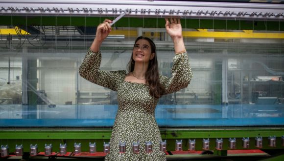
Researchers from Tel Aviv University used CRISPR to edit thousands of genes in tomato plants.

Researchers from the School of Plant Sciences and Food Security at the Wise Faculty of Life Sciences at Tel Aviv University have developed a genetic editing method tailored to crop plants, which has influenced various traits in tomato plants, including the taste and shape of the fruit. The researchers believe this innovative technology can be applied to various crop species and may eventually be used to cultivate new and improved plant varieties. “We demonstrated that with our technology, it is possible to select specific traits and influence them, a capability that is essential for advancing agriculture and achieving food security,” the researchers stated.

The study was led by Prof. Eilon Shani, Prof. Itay Mayrose and PhD student Amichai Berman (School of Plant Sciences and Food Security at Tel Aviv University) together with PhD student Ning Su and Dr. Yuqin Zhang (University of Chinese Academy of Sciences in Beijing), and Dr. Osnat Yanai from the Israeli Agri-Tech company NetaGenomiX. The article was published in the prestigious journal Nature Communications.

Prof. Shani explains: “Researchers around the world are engaged in advancing agriculture in order to address accelerated global changes and feed the global population in the coming decades. Among other things, genetic editing technologies are being advanced to develop new plant varieties with desirable traits such as resistance to drought, heat, and disease, improved flavor, optimized nutrient usage, and more. One such method is CRISPR-Cas9, which has revolutionized the field of genetic editing by enabling the precise modification of specific genes in the genome.

However, in the realm of agricultural development, this method has encountered several fundamental challenges: Firstly, while CRISPR technology allows for targeted gene editing, until now, this capability was limited in scale – the number of genes that could be edited and studied was very small. In the current study, we significantly improved the method’s efficiency, enabling us to examine the roles of thousands of genes. Secondly, many plants exhibit ‘genetic redundancy’: different genes from the same family, composed of similar amino acid sequences, compensate for one another and preserve the trait even if one gene is deactivated or edited”.

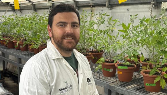
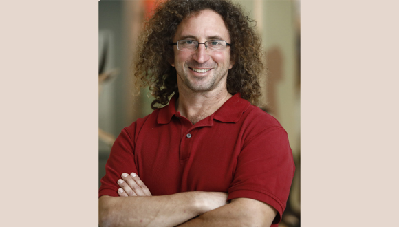
PhD student Amichai Berman.

Amichai Berman: “To overcome genetic redundancy, we aimed to alter entire families of similar genes simultaneously. In a previous study, we developed a breakthrough solution to overcome the issue of genetic redundancy, a dedicated algorithm, and fed it a list of thousands of genes we wanted to edit. The algorithm identified a suitable CRISPR unit for each gene (or gene group) on the list that would induce the desired modification, thereby constructing CRISPR libraries. The first study achieved good results in the model plant Arabidopsis thaliana, and this time we sought to test the method in a crop plant for the first time. We chose the tomato.”

In the current study, the researchers built 10 libraries comprising approximately 15,000 unique CRISPR units targeting the tomato genome – each unit designed to affect a specific gene group from the same family. They then used the CRISPR units to induce mutations in around 1,300 tomato plants, each plant with an alteration in a different gene group. The researchers then tracked the development of each plant to examine whether the selected changes appeared in fruit size, shape, taste, nutrient utilization, or pest resistance. Indeed, the researchers identified several lines with sweetness levels either lower or higher than the control plants.

Prof. Shani concludes: “In this study, using our innovative method, we successfully made targeted genetic changes to gene families in the tomato plant, and identified precisely which genetic edits produced the desired result.” The Israeli Agri-Tech company NetaGenomiX has received a license to commercialize the new technology, with the goal of advancing food security by developing non-GMO crops adapted to the changing climate, providing benefits for both farmers and consumers.

Amichai Berman adds: “We believe our research opens the door to breeding improved varieties for a wide range of crops and also advances the field of plant science as a whole. In follow-up studies, we are working on developing additional selected traits in tomatoes and in rice.”

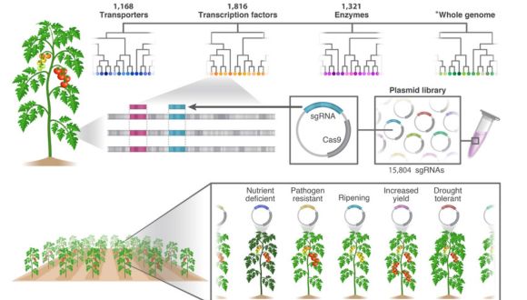
Large-Scale Functional Genomics in Tomato Using a High-Throughput Multi-targeted CRISPR Screening Approach. The tomato plant genome is divided into gene families. For each group of similar genes, a unique CRISPR unit is designed to alter their function (in total, over 15,000 CRISPR units were designed). These CRISPR units are delivered into tomato plants, which are then monitored for growth and development. In the final stage, plants exhibiting changes in selected traits are identified and genetically and physiologically characterized. This new approach enables the large-scale targeting of genetic redundancy within gene families, on the scale of hundreds of genes.

Unlocking Quantum Mysteries with Pendula

Pendulum Experiment Sheds Light on Quantum Mysteries in Topological Materials, Revealing Insights Unreachable by Traditional Methods

A recent study conducted at Tel Aviv University has devised a large mechanical system that operates under dynamical rules akin to those found in quantum systems. The dynamics of quantum systems, composed of microscopic particles like atoms or electrons, are notoriously difficult, if not impossible, to observe directly. However, this new system allows researchers to visualize phenomena occurring in specialized “topological” materials through the movement of a system of coupled pendula.

The research is a collaboration between Dr. Izhar Neder of the Soreq Nuclear Research Center, Chaviva Sirote-Katz of the Department of Biomedical Engineering, Dr. Meital Geva and Prof. Yair Shokef of the School of Mechanical Engineering, and Prof. Yoav Lahini and Prof. Roni Ilan of the School of Physics and Astronomy at Tel Aviv University and was recently published in the Proceedings of the National Academy of Sciences of the USA (PNAS)

Exploring Quantum Wave Phenomena

Quantum mechanics governs the microscopic world of electrons, atoms and molecules. An electron, which is a particle that moves in an atom or in a solid, may have properties that give rise to wave-like phenomena. For instance, it may demonstrate a probability of dispersing in space similar to waves spreading out in a pool after a stone is thrown in, or the capability to exist simultaneously in more than one place.

Such wave-like properties lead to a unique phenomenon that appears in some solid isolators, where even though there is no electric current through them, and the electrons do not move due to an external electric voltage, the internal arrangement of the material shows up in a state referred to as “topological”. This means that the wave of electrons possesses a quantity that can “close on itself” in different ways, somewhat like the difference between a cylinder and a Möbius strip. This “topological” state of the electrons, for which the 2016 Nobel Prize in Physics was awarded, is considered a new state of matter and attracts much current research.

Chaviva Sirote-Katz

Despite the theoretical interest, there is a limitation in measuring these phenomena in quantum systems. Due to the nature of quantum mechanics, one cannot directly measure the electron’s wave function and its dynamical evolution. Instead, researchers indirectly measure the wave-like and topological properties of electrons in materials, for instance by measuring the electrical conductivity at the edges of solids.

In the current study, the researchers considered the possibility of constructing a sufficiently large mechanical system that would adhere to dynamical rules akin to those found in quantum systems, and in which they could directly measure everything. To this end, they built an array of 50 pendula, with string lengths that slightly varied from one pendulum to the other. The strings of each neighboring pair of pendula were connected at a controlled height, such that each one’s motion would affect its neighbors’ motion.

Quantum Pendulum Insights

On one hand, the system obeyed Newton’s laws of motion, which govern the physics of our everyday lives, but the precise lengths of the pendula and the connections between them created a magical phenomenon: Newton’s laws caused the wave of the pendulum’s motion to approximately obey Schrödinger’s equation – the fundamental equation of quantum mechanics, which governs the motion of electrons in atoms and in solids. Therefore, the motion of the pendula, which is visible in the macroscopic world, reproduced the behaviors of electrons in periodic systems such as crystals.

The researchers pushed a few pendula and then released them. This generated a wave that propagated freely along the chain of pendula, and the researchers could directly measure the evolution of this wave – an impossible mission for the motion of electrons. This enabled the direct measurement of three phenomena. The first phenomenon, known as Bloch oscillations, occurs when electrons within a crystal are influenced by an electric voltage, pulling them in a specific direction. In contrast to what one would expect, the electrons do not simply move along the direction of the field, but they oscillate back and forth due to the periodic structure of the crystal. This phenomenon is predicted to appear in ultra-clean solids, which are very hard to find in nature. In the pendula system, the wave periodically moved back and forth, exactly according to Bloch’s prediction.

The second phenomenon that was directly measured in the pendula system is called Zener tunneling. Tunneling is a unique quantum phenomenon, which allows particles to pass through barriers, in contrast to classical intuition. For Zener tunneling, this appears as the splitting of a wave, the two parts of which then move in opposite directions. One part of the wave returns as in Bloch oscillations, while the other part “tunnels” through a forbidden state and proceeds in its propagation. This splitting, and specifically its connection to the motion of the wave in either direction, is a clear characteristic of the Schrödinger equation.

In fact, such a phenomenon is what disturbed Schrödinger, and is the main reason for the suggestion of his famous paradox; according to Schrödinger’s equation, the wave of an entire cat can split between a live-cat state and a dead-cat state. The researchers analyzed the pendula motion and extracted the parameters of the dynamics, for instance, the ratio between the amplitudes of the two parts of the split wave, which is equivalent to the quantum Zener tunneling probability. The experimental results showed fantastic agreement with the predictions of Schrödinger’s equation.

The pendula system is governed by classical physics. Therefore, it cannot mimic the full richness of quantum systems. For instance, in quantum systems, the measurement can influence the system’s behavior (and cause Schrödinger’s cat to eventually be dead or alive when it is viewed). In the classical system of macroscopic pendulum, there is no counterpart to this phenomenon. However, even with these limitations, the pendula array allows the observation of interesting and non-trivial properties of quantum systems, which may not be directly measured in the latter.

The third phenomenon that was directly observed in the pendula experiment was the wave evolution in a topological medium. Here, the researchers found a way to directly measure the topological characteristic from the wave dynamics in the system – a task that is almost impossible in quantum materials. To this end, the pendula array was tuned twice, so that they would mimic Schrödinger’s equation of the electrons, once in a topological state and once in a trivial (i.e. standard) state. By comparing small differences in the pendulum motion between the two experiments, the researchers could classify the two states. The classification required a very delicate measurement of a difference between the two experiments of exactly half a period of oscillation of a single pendulum after 400 full oscillations that lasted 12 minutes. This small difference was found to be consistent with the theoretical prediction.

The experiment opens the door to realizing further situations that are even more interesting and complex, like the effects of noise and impurities, or how energy leakage affects wave dynamics in Schrödinger’s equation. These are effects that can be easily realized and seen in this system, by deliberately perturbing the pendula motion in a controlled manner.

The Bat’s Walking Cane: How a Tail Becomes a Sensor in the Dark

New research highlights a unique evolutionary adaptation: a bat’s tail acting as a reverse walking cane.

A new study from Tel Aviv University reveals that the Greater Mouse-Tailed Bat (Rhinopoma microphyllum) uses its long tail as a natural tactile sensor to navigate backward in dark caves. The researchers discovered that the bat’s long tail functions as a dynamic sensing tool, enabling it to avoid obstacles and orient itself in complex environments while climbing backward—especially when other sensory abilities like vision and echolocation are limited. The study found that when the tail was numbed with a local anesthetic, the bats’ ability to navigate around obstacles while crawling backward and the speed of their movement decreased significantly.

The research was led by Sahar Hajyahia and Mor Taub, students in the laboratory of Prof. Yossi Yovel of Tel Aviv University’s School of Zoology in the Wise Faculty of Life Sciences and the Sagol School of Neuroscience. The study was published in the scientific journal iScience.

פרופ' יוסי יובל

Prof. Yossi Yovel.

Professor Yossi Yovel explains: “During the study, the bats crawled up a vertical maze while moving backward as we recorded their movement using an advanced tracking system. The bats used their tails like a blind walking cane, swinging them from side to side to detect obstacles and climb more safely and efficiently. In contrast, when the tail was numbed, the climbing time increased by an average of 10%, and the bats made more lateral movements instead of moving upward, apparently trying to find their way”.

The researchers also noted that the bats demonstrated a remarkable ability to distinguish between different textures using their tails. They were able to differentiate between a fine wooden grid (1 cm intervals) and a sparser grid (1.5 cm), highlighting the tail’s complex ability to serve as an exceptionally sensitive tactile sensor.

Greater Mouse-Tailed Bat (Photo courtesy of Jens Rydell).

Professor Yossi Yovel concludes: “In most bats, the tail is very short and integrated into the wing membrane. In Rhinopoma, however, the tail remains long and free, and to the best of our knowledge, they are the only bats that use it to sense their immediate surroundings. This is another example of how evolution adapts animals’ senses to meet specific needs—in this case, moving backward in dark places around obstacles and other bats. Many bats crawl backward on dark walls and cannot use their frontal senses like vision and sonar to ‘see’ behind them. One can think of the tail as a sort of reverse sensor for the Rhinopoma. This discovery opens the door to further research on tail usage as a sensor in other animal species. Additionally, the findings could inspire the development of new sensory technologies inspired by nature, such as robotic navigation systems for complex environments”.

Why Do City Bats Give Birth Earlier?

Research reveals the city’s surprising effect on birth timing.

A groundbreaking study from Tel Aviv University, the first of its kind on mammals, has found that bats living in urban environments give birth, on average, about 2.5 weeks earlier than bats living in rural areas. The researchers attribute this difference in birthing times between the city and the countryside to more favorable temperatures and greater food abundance in urban areas. Bats are mammals, making this the first study to link the urban living environment to the timing of birth in mammals.

The research was led by the lab team of Prof. Yossi Yovel from the School of Zoology in the Wise Faculty of Life Sciences and the Steinhardt Museum of Natural History at Tel Aviv University. The study included contributions from Dr. Maya Weinberg, Dean Zigdon, and Mor Taub, and was published in the scientific journal BMC Biology.

פרופ' יוסי יובל

Prof. Yossi Yovel.

Over three years, the researchers monitored ten bat colonies in urban and rural areas, sampling hundreds of female bats and approximately 120 pups from these colonies. The findings revealed that pups born in urban colonies were, on average, 2.5 weeks older, as evidenced by their longer forearms and higher body weights compared to pups from rural colonies.

Prof. Yovel explains: “Fruit bats living in cities benefit from favorable environmental conditions, including higher temperatures due to the ‘urban heat island’ effect and greater food availability, primarily from ornamental fruit trees irrigated year-round. These conditions allow urban bats to cope better with harsh winters and start their reproductive cycles earlier. This enables females to give birth earlier in the season, increasing their chances of becoming pregnant again within the same year.”

However, the researchers emphasize that it remains unclear whether the bats are shortening their pregnancies (a capability known in some bat species) or becoming pregnant earlier. They add that the study opens the door to further research on how urbanization affects mammalian reproductive patterns in general and bats in particular, and how these findings can be used to protect other species in changing ecosystems. “This study highlights the importance of understanding the connection between animals and their environments, especially in an era when urbanization is reshaping the planet,” concludes Prof. Yovel.

A mother bat in flight, carrying her pup beneath her in the city and countryside (Photo credit: Yuval Barkai).

City Life Keeps These Birds on the Move

City birds roam, rural birds stay put—how urban life shapes movement.

Researchers from Tel Aviv University’s School of Zoology studied the behavior and movement of the common spur-winged lapwing. They compared the habits of “urban lapwings” to those of their “rural” counterparts. In this latest study — the third in a series — the researchers tracked spur-winged lapwings in the Beit She’an and Harod Valleys, finding that birds living in human settlements exhibited greater mobility than those in nearby fishponds and fields, especially during the nesting season.

The researchers note: “We often see birds in populated areas and cities, demonstrating that some species habituate and even flourish in human environments. However, we tend to overlook how human presence affects these animals, altering their behavior — much like the classic story of the ‘town mouse and country mouse”.

The study was led by doctoral student Michael Bar-Ziv from the School of Zoology under the guidance of Prof. Orr Spiegel. Additional contributors included Hilla Ziv, Mookie Breuer, Eitam Arnon, and Assaf Uzan, all members of the Movement Ecology and Individual Behavior Laboratory at the School of Zoology from The George S. Wise Faculty of Life Sciences at Tel Aviv University.

פרופ' אור שפיגל

Prof. Orr Spiegel.

Prof. Spiegel explains: “Our lab focuses on animal behavior and movement in space, aiming to conserve nature and prevent zoonotic disease transmission. In our latest series of studies on this system, we sought to examine how urbanization and human presence influence animals, particularly birds. We chose to focus on the spur-winged lapwing (Vanellus spinosus). These common species are often found in populated areas and are large enough to carry a transmitter that continuously tracks its location. In our two earlier studies, we found that urban lapwings are bolder than their counterparts that live in more natural areas and that these behavioral differences persist even when tested in a shared and controlled environment, such as aviaries at the zoological garden at Tel Aviv University”.

How Habitat Shapes Bird Movement

This study focused on spur-winged lapwings living in the Beit She’an and Harod Valleys, comparing the spatial movement patterns of two adjacent sub-populations: those living in moshavim and kibbutzim and those in nearby fishponds and fields. Two main parameters were examined: mobility — the daily distance traveled, and exploration — whether they revisit familiar locations or explore new, unfamiliar sites.

הי? אתם מחפשים אותי? סיקסק סקרן (צילום: היל זיו)

Close-up of a Spur-winged Lapwing (Photo creditHilla Ziv).

Michael Bar-Ziv explains: “Spur-winged lapwings nest during spring, building their nests on the ground. We captured them at their nests, attached transmitters, and released them to continue their lives. The transmitters, which sent a signal every eight seconds, allowed us to track the birds over several months”. In total, the researchers tracked 135 individuals — half males and half females — with an average tracking period of 194 days. The longest tracking period lasted about ten months. The data collected underwent statistical analysis to compare the movement and behavioral patterns of the two groups.

Bar-Ziv adds: “First, we found that each bird has its own distinct personality, reflected in consistent movement patterns that differ from those of other individuals. Additionally, we discovered that lapwings living in human settlements are more mobile than those in undeveloped areas. We believe this is because disturbances in towns and villages cause the birds to take flight more often, increasing their overall flight distance but not their maximum distance from the nest. As for exploration, we found a more complex picture, with seasonal differences and variations between males and females. For example, during the nesting season, females in fields were more exploratory than their counterparts in populated areas, while the opposite was true for males. We don’t yet have an explanation for these patterns”.

Bird City Life

Prof. Spiegel concludes: “Many animals live around us in cities and towns, and some species even adapt well to human environments. However, research shows that even among these city-dweller species, various aspects of behavior change due to human presence compared to their behavior in the wild. Alternatively, individuals with certain personality traits may be more likely to choose urban living, which explains behavioral differences. In this study, we found significant differences, particularly increased mobility among ‘urban lapwings’ during the nesting season. Future studies will explore the potential influence of urban environments on the development and personality of chicks from a young age to adulthood. These studies will help us distinguish between the mechanisms contributing to the observed differences — whether certain types of individuals gravitate toward cities or whether urban environments shape behavior”.

Is the Spread of Deadly Pathogens Threatening Coral Reefs Worldwide?

From Eilat to the Indian Ocean, a relentless pathogen is ravaging marine ecosystems

An international team of researchers, led by scientists from Tel Aviv University, has discovered that the pathogen responsible for the mass deaths of sea urchins along the Red Sea coast is the same one responsible for mass mortality events among sea urchins off the coast of Réunion Island in the Indian Ocean. This finding raises fears that the pathogen – a waterborne ciliate – could spread further, to the Pacific Ocean. The researchers warn that this is a highly aggressive global pandemic, and are now spearheading an international effort to track the disease and preserve sea urchins, which play a crucial role in the health of coral reefs.

The study, led by Dr. Omri Bronstein from the School of Zoology at Tel Aviv University’s Wise Faculty of Life Sciences and the Steinhardt Museum of Natural History, was published in the prestigious journal Ecology.

The Research Team. Photo credit: Tel Aviv University.

“This is a first-rate ecological disaster”, explains Dr. Bronstein.

She continues: “Sea urchins are vital to the health of coral reefs. They act as the ‘gardeners’ of the reef by feeding on algae, preventing it from overgrowing and suffocating the coral, which competes with algae for sunlight. In 1983, a mysterious disease wiped out most of the Diadema sea urchins in the Caribbean. Unchecked, the algae there proliferated, blocking sunlight from the coral, and the region shifted from a coral reef ecosystem to an algae-dominated one. Even 40 years later, the sea urchin population — and consequently the reef — has not recovered”.

In 2022, the disease reemerged in the Caribbean, targeting the surviving sea urchin populations and individuals. This time, armed with advanced scientific and technological tools to collect and interpret the forensic evidence, researchers at Cornell University successfully identified the pathogen as a ciliate Scuticociliate parasite. A year later, in early 2023, Dr. Bronstein was the first to identify mass mortality events among long-spined sea urchins, a close relative of the Caribbean Sea urchins, in the Red Sea.

Sea urchin mortalities on Reunion Island. Photo credit: Jean-Pascal Quod.

“Until recently, this was one of the most common sea urchins in Eilat’s coral reefs — the familiar black urchins with long spines,” says Dr. Bronstein. “Today, this species no longer exists in significant numbers in the Red Sea. The event was extremely violent: within less than 48 hours, a healthy population of sea urchins turned into crumbling skeletons. In some locations in Eilat and the Sinai, mortality rates reached 100 percent. In follow-up research, we demonstrated that the Caribbean pathogen was the same one affecting populations in the Red Sea”.

Genetic Proof Links Global Sea Urchin Deaths to One Pathogen

Now, using genetic tools, Dr. Bronstein and his international colleagues have shown that the same ciliate parasite is responsible for similar mortality events off the coast of Réunion Island in the Indian Ocean. “This is the first genetic confirmation that the same pathogen is present in all these locations,” he says. “Now it’s a global event, a pandemic. The Caribbean, Red Sea, and the Indian Ocean are critical regions for the world’s coral reefs, and mortality rates for sea urchins in these areas are very high — over 90 percent. As of now, we have no evidence of this pathogen in Pacific Ocean sea urchins, but this is something we are actively investigating. Although we’ve developed genetic tools for the specific identification of the pathogen, it’s difficult to monitor such rapid extinction events in the vast underwater environment. We are terrestrial creatures, and some reefs are located in deep or remote areas. If we miss the mortality event by even a couple of days, we might find no trace of the extinct population”.

 Four healthy sea urchin species on Reunion Island. Photo credit: Jean-Pascal Quod.

To track the progression of the pandemic, Dr. Bronstein has established an international network of collaborators. He provides them with alerts about the likelihood of mortality events in their regions and sends them the necessary equipment to sample and preserve affected sea urchins for comparison with samples from other locations. These kits are then sent back to the laboratory at Tel Aviv University.

“For populations that are already infected, we really have no tools to help them,” says Dr. Bronstein with regret. “There is no Pfizer or Moderna for sea urchins — not because we don’t want one, but because we simply can’t treat them underwater. Our focus must be on two entirely different tracks. The first is prevention. To prevent further spread of the pandemic, we need to understand why it erupted here and now. We’ve developed two hypotheses for this. The first is the transportation hypothesis — that the pathogen from the Caribbean was transported by humans to new and distant regions after being carried in the ballast water of ships, infecting sea urchins in the Red Sea before spreading to the Western Indian Ocean.

The sea urchin Diadema setosum before (left) and after (right) mortality. The white skeleton is exposed following tissue disintegration and loss of spines. Photo credit: Tel Aviv University.

How climate change might be spreading marine diseases worldwide

Incidentally, if this hypothesis is correct, we would expect to see mortality events in West Africa as well — as many cargo ships from the Caribbean stop there on their way to the Mediterranean and then through the Suez Canal to the Red Sea. Indeed, just in the past few weeks, we’ve discovered widespread mortality events in West Africa, as we predicted, and we’ve managed to obtain a limited number of samples collected during these events, which we are currently analyzing in the lab. If ships are indeed the source of the spread, then we could think of mitigation strategies. It’s not simple, and ships will never be completely sterile, but there are measures we can take. The second possibility is even more concerning: that the pathogen has always been present, and climatic changes have triggered its virulence and outbreak. That’s a challenge of an entirely different magnitude, one that we, as marine biologists, have very limited means to address”.

In parallel with global efforts, Dr. Bronstein has recently established a breeding nucleus for the affected sea urchins at the Israel Aquarium in Jerusalem, in collaboration with the Biblical Zoo and the Israel Nature and Parks Authority. This breeding population will serve as a reserve to restore affected populations and advance research into infection mechanisms and possible treatments.

“The pathogen is transmitted through water, so even sea urchins raised for research purposes in aquariums at the Interuniversity Institute for Marine Sciences and the Underwater Observatory in Eilat became infected and died. That is why we established a breeding nucleus with the Israel Aquarium, whose aquariums are completely disconnected from seawater. We genetically test the urchins transferred to the nucleus to ensure they are not carriers of the disease and that they genetically belong to the Red Sea population, enabling us to rehabilitate the population in the future. At the same time, we are using them to develop sensitive genetic tools for early disease detection from seawater samples — essentially creating ‘underwater COVID tests’ for sea urchins”.


Can Foreign DNA Outsmart Bacterial Defenses?

A step toward combating resistance and manipulating bacteria

A new study by Tel Aviv University reveals how bacterial defense mechanisms can be neutralized, enabling the efficient transfer of genetic material between bacteria. The researchers believe this discovery could pave the way for developing tools to address the antibiotic resistance crisis and promote more effective genetic manipulation methods for medical, industrial, and environmental purposes. The study was led by PhD student Bruria Samuel from the lab of Prof. David Burstein at the Shmunis School of Biomedicine and Cancer Research at Tel Aviv University’s Wise Faculty of Life Sciences. Other contributors to the research include Dr. Karin Mittelman, Shirly Croitoru, and Maya Ben-Haim from Prof. Burstein’s lab. The findings were published in the prestigious journal Nature.

The researchers explain that genetic diversity is essential for the survival and adaptation of different species in response to environmental changes. For humans and many other organisms, sexual reproduction is the primary driver of the genetic diversity required for survival. However, bacteria and other microorganisms lack such a reproduction mechanism. Nevertheless, as demonstrated by the alarming speed at which antibiotic resistance spreads among bacterial populations, bacteria have alternative mechanisms to maintain the genetic diversity necessary for survival, including the direct DNA transfer between bacteria.

DNA transfer between bacteria plays a crucial role in their survival. Yet, a key aspect of this process has remained underexplored: how is the exchange of genetic material so prevalent despite bacteria having a wide range of defense mechanisms designed to destroy any foreign genetic material entering their cells? The new research focuses on “conjugation”, one of the main mechanisms for transferring DNA from one bacterium to another. During conjugation, one bacterial cell connects directly to another through a tiny tube that allows the transfer of genetic material fragments known as plasmids. Prof. Burstein explains: “Plasmids are small, circular, double-stranded DNA molecules classified as ‘mobile genetic elements.’ Like viruses, plasmids move from one cell to another, but unlike viruses, they do not need to kill the host bacterium to complete the transfer”.

Plasmids That Outsmart Bacterial Defenses

As part of the natural exchange, plasmids often give recipient bacteria genetic advantages. For example, many antibiotic-resistance genes spread through plasmid transfer between bacteria. However, bacteria also have numerous defense mechanisms aimed at eliminating any foreign DNA entering their cells. “Conjugation is a well-known process that scientists also use in the lab to transfer genes between bacteria. It’s also known that bacteria possess mechanisms to destroy foreign DNA, including plasmid DNA, and some of these mechanisms are even used for various research purposes. However, until now, no one has fully explored how plasmids overcome these defense mechanisms”, says Prof. Burstein. Samuel explains that she began the research by conducting a computational analysis of 33,000 plasmids and identifying genes associated with ‘anti-defense’ systems that help plasmids bypass bacterial defense mechanisms. What was even more interesting was the location of these genes. As mentioned, plasmids are double-stranded circular DNA segments. To pass through the thin tube that connects the bacteria, one of those circular strands is cut at a certain point by a protein, which then binds to the cleaved strand and initiates its transfer to the recipient cell. “The genes for the anti-defense systems that I identified were found to be concentrated near that cutting point, and organized in such a manner that they would be the first genes to enter the new cell. This strategic positioning allows the genes to be activated immediately upon transfer, giving the plasmid the advantage needed to neutralize the recipient bacteria’s defense systems”.

Left to right: Prof. David Burstein & PhD student Bruria Samuel.

Prof. Burstein recounts how, when Samuel first showed him her results, he found it hard to believe that such a phenomenon had not been identified before. “Bruria conducted an extensive literature review and found that no one had previously made this connection,” he says. Since the discovery was made by analyzing existing databases with computational tools, the next step was to demonstrate in the lab that this phenomenon indeed occurs during plasmid transfer between bacteria. Samuel explains, “To do this, we used plasmids that confer antibiotic resistance and introduced them into bacteria equipped with CRISPR, the well-known bacterial defense system that can target and destroy DNA, including that of plasmids. This method allowed us to easily test the conditions under which the plasmid could overcome the defense system — if it succeeds in overcoming the CRISPR system, the recipient bacteria become resistant to antibiotics. If it fails, the bacteria die”. Using this method, Samuel demonstrated that if the anti-defense genes are positioned near the DNA entry point, the plasmid successfully overcomes the CRISPR system. However, if these genes are located elsewhere on the plasmid, the CRISPR system destroys the plasmid, and the bacteria die upon exposure to antibiotics.

How Can Gene Transfers Be Improved?

Prof. Burstein notes that understanding the positioning of anti-defense systems on plasmids could enable the identification of new anti-defense genes, a subject currently under highly active research. “Moreover, our study can contribute to designing more efficient plasmids for genetic manipulation of bacteria in industrial processes. While plasmids are already widely used for these purposes, the efficiency of plasmid-based genetic transfer in lab conditions is significantly lower than that of natural plasmids,” he says. “Another potential application could involve designing effective plasmids for genetic manipulation of natural bacterial populations. This could help block antibiotic resistance genes in hospital bacterial populations, teach bacteria in soil and water to break down pollutants or fix carbon dioxide, and even manipulate gut bacteria to improve human health”.

Ramot, Tel Aviv University’s technology transfer company, regards this discovery as a significant biotechnological breakthrough with broad applications. Dr. Ronen Kreizman, CEO of Ramot, states: “First, I want to congratulate Prof. David Burstein and his lab team on this fascinating scientific discovery. The new research opens revolutionary possibilities in areas such as developing drugs against resistant bacteria, synthetic biology, agritech and foodtech. The ability to control and fine-tune genetic material transfer between bacteria could become a powerful tool for addressing environmental, agricultural, and medical challenges. We are currently working on commercializing this technology to realize its full potential”.

TAU Discovery Decodes a Rare Neurological Disease

This breakthrough could pave the way for neurological treatments.

Researchers at Tel Aviv University have developed an innovative research model that allowed them to decode the mechanism underlying a severe and rare neurological disease. The disease is characterized by symptoms such as epilepsy, developmental delay, and intellectual disability.

According to the researchers: “Decoding the disease mechanism is a critical step toward developing treatments targeting specific cellular functions for this disease and other conditions with similar mechanisms affecting cellular energy production”.

The research was led by Tel Aviv University’s Prof. Abdussalam Azem, Dean of the Wise Faculty of Life Sciences, in collaboration with Prof. Uri Ashery and PhD student Eyal Paz from the School of Neurobiology, Biochemistry and Biophysics at the Wise Faculty of Life Sciences and the Sagol School of Neuroscience. Additional contributors included Dr. Sahil Jain and Dr. Irit Gottfried from the School of Neurobiology, Biochemistry, and Biophysics at Tel Aviv University, Dr. Orna Staretz-Chacham from the Faculty of Health Sciences at Ben-Gurion University, Dr. Muhammad Mahajnah from the Technion, and researchers from Emory University in Atlanta, USA. The findings were published in the prominent journal eLife.

TIMM50 Mutation Linked to Rare Brain Disorders

Prof. Azem explains: “The disease we studied is caused by a mutation in a protein called TIMM50, which plays a crucial role in importing other proteins into the mitochondria—the organelle considered the cell’s energy powerhouse. The human mitochondria operate with about 1,500 proteins (approximately 10% of all human proteins), but only about 13 of them are produced within the mitochondria itself. The rest are imported externally through various mechanisms. In recent years, mutations in the TIMM50 protein, which is responsible for importing about 800 proteins into the mitochondria, were found to cause severe and rare neurological disease with symptoms like epilepsy, developmental delay, and intellectual disability”.

Prof. Ashery adds: “Protein import into the mitochondria has been extensively studied over the years, but how a mutation in TIMM50 affects brain cells was never tested before. To investigate this for the first time, we created an innovative model using mouse neurons that mimics the disease caused by the TIMM50 protein mutation. In this study, we significantly reduced the expression of the protein in mouse brain cells and observed its impact on the cells”.

How Does a Protein Defect Link Energy Loss to Epilepsy?

Eyal Paz explains: “The impairment of the protein led to two main findings: a reduction in energy production in the neurons, which could explain the developmental issues seen in the disease and an increase in the frequency of action potentials (the electrical signals that transmit information along neurons and enable communication between them). This increase in action potential frequency is known to be associated with epilepsy. The change in frequency is likely caused by significant damage to two proteins that function as potassium channels. Imbalances in potassium levels can lead to life-threatening conditions, such as arrhythmias, cardiac arrest, and muscle weakness, potentially leading to paralysis. These potassium channels may serve as potential targets for future drug treatments for the disease”.

Prof. Azem concludes: “Our study decodes the mechanism of a severe and rare neurological disease caused by a mutation in a protein critical for importing proteins into the mitochondria. Understanding the mechanism is a crucial step toward treatment, as it enables the development of drugs targeting the specific issues identified. Additionally, we created a new research model based on mouse neurons that significantly advances the study of protein import into mitochondria in brain cells. We believe that our findings, combined with the innovative model, will enable more in-depth research and the development of treatments for various neurological diseases caused by similar mitochondrial dysfunction mechanisms”.

What Can Locusts Teach Us About Efficiency in Design?

Research shows locusts’ digging valves are built just right for their task.

Researchers at Tel Aviv University examined the mechanical wear of digging valves located at the tip of the female locust’s abdomen, used to dig pits for laying eggs 3 to 4 times during her lifetime. They found that, unlike organs with remarkably high wear resistance, such as the mandible (lower jaw), the valves wear down substantially due to intensive digging.

The researchers: “This is an instructive example of the ‘good enough’ principle in nature. Evolution saw no need to invest extra energy and resources in an organ with a specific purpose that performs its function adequately. We, humans, who often invest excessive resources in engineered systems, can learn much from nature”.

The study was led by Dr. Bat-El Pinchasik from the School of Mechanical Engineering and Prof. Amir Ayali from the School of Zoology at the Wise Faculty of Life Sciences, the Sagol School of Neuroscience and the Steinhardt Museum of Natural History at Tel Aviv University. Other participants included: PhD student Shai Sonnenreich from TAU’s School of Mechanical Engineering, as well as researchers from the Technical University of Dresden in Germany, Prof. Yael Politi and a postdoc in her group, Dr. Andre Eccel Vellwock. The article was published in the prestigious journal Advanced Functional Materials.

Left to right: Prof. Amir Ayali, Dr. Bat-El Pinchasik & PhD student Shai Sonnenreich.

Dr. Pinchasik: “In my lab, we study mechanical mechanisms in nature, partly to draw inspiration for solving technological problems. Recently we collaborated with locust expert Prof. Amir Ayali in a series of studies, to understand the mechanism used by the female locust for digging a pit to lay her eggs. This unique mechanism consists of two shovel-like valves that open and close cyclically, digging into the soil while pressing the sand against the walls”.

Prof. Ayali: “We know that many mechanisms in the bodies of insects in general, and locusts in particular, are exceptionally resistant to mechanical wear. For example, the locust’s mandibles, used daily for feeding, are made of a highly durable material. The digging valves, on the other hand, while subjected to substantial shear forces during digging, are used only 3 or 4 times in the female’s lifetime – when she lays eggs. In this study, we sought to discover whether these digging valves, made of hard cuticular material, were also equipped by evolution with high resistance to mechanical wear”.

To address this question, the researchers examined the digging valves in three different groups of female locusts: young females that had not yet laid eggs, mature females kept in conditions that prevented them from laying eggs – to test whether age alone causes wear and adult females that had already laid eggs 3 or 4 times. To analyze the internal structure and durability of the digging valves, the researchers used several advanced technologies: confocal microscopy, 3D fluorescent imaging, and a particle accelerator (synchrotron) in collaboration with the German team. The findings indicated significant signs of wear in the valves and a lack of elements associated with high resistance to mechanical wear. Notably, no reinforcing metal ions, typical of extremely wear-resistant biological materials, were found in the valves.

Dr. Pinchasik: “A female locust’s biological role is laying eggs three or four times in her life. In this study, we found that evolution has designed her digging valves to meet their task precisely—no more and no less. This is a wonderful example of the ‘good enough’ principle in nature: no extra resources are invested in an organ when they’re not needed”.

“As humans, we can learn much from nature – about conserving materials, energy, and resources. As engineers who develop products, we must understand the need precisely and design an accurate response, avoiding unnecessary overengineering” – Dr. Pinchasik.

Eyes Wide Shut: Bats Can Navigate Long Distances Using Sound Alone

Researchers found that bats can create a mental “sound map” of their environment.

A new study by Tel Aviv University and the Steinhardt Museum of Natural History has proven, for the first time, that bats can navigate in nature over many kilometers using only echolocation, without relying on other senses. The researchers explain: “It’s well-known that bats are equipped with a natural sonar, allowing them to emit sound waves that bounce back from nearby objects, helping them navigate. However, it’s also known that bats use their sense of sight during flight. Laboratory studies have shown that bats can navigate within enclosed spaces using only echolocation — but sonar ‘sees’ only about 10 meters ahead, so what happens under natural conditions, in open areas stretching over many kilometers? Can bats rely solely on echolocation for long-distance navigation?” In this study, that question was explored in depth for the first time.

They Follow the Echo

The research was led by Prof. Yossi Yovel of Tel Aviv University’s School of Zoology, Sagol School of Neuroscience, and Steinhardt Museum of Natural History, along with Dr. Aya Goldshtein, formerly a doctoral student of Prof. Yovel and currently a researcher at the Max Planck Institute in Germany. Additional partners from Tel Aviv University included Prof. Sivan Toledo of the Blavatnik School of Computer Science; Xing Chen, Dr. Eran Amichai, and Dr. Arjan Boonman of the School of Zoology; and Lee Harten of the Sagol School of Neuroscience. Prof. Ran Nathan and Dr. Yotam Orchan of the Hebrew University and Prof. Iain Couzin of the Max Planck Institute in Germany also participated in the study, which was published in the journal Science.

The innovative research carried out over six years, utilized a unique tracking system installed in Israel’s Hula Valley. Using this GPS-like technology, the researchers could track the flight of tiny bats from the species known as Kuhl’s pipistrelle, each weighing only six grams —— the smallest mammal ever to be monitored in this way.

For the study, the researchers collected around 60 bats from their roost in the Hula Valley area and moved them about three kilometers away from the roost — still within their familiar habitat. A tag was attached to each bat, and the eyes of some were covered with a cloth strip, temporarily preventing them from seeing during flight, though they could remove the covering with their feet upon landing. In addition, the researchers employed techniques to temporarily disrupt the bats’ sense of smell and magnetic sense, thereby creating conditions in which they would be able to find their way home using only echolocation. Remarkably, the bats managed to return to their roost without difficulty.

In the second phase, the researchers built a computerized acoustic model of the bats’ natural environment in the Hula Valley. Prof. Yovel explains: “This model is based on a 3D map of the area where the bats navigate, reflecting the echoes that the bat hears as it uses echolocation to journey through its surroundings. In examining the bats’ flight paths, we discovered that they choose routes where the echoes contain a lot of information, which helps them navigate. For example, an area rich in ​​vegetation, such as bushes and trees, returns echoes with more information than an open field, making bats less likely to fly over open terrain. We also found that some areas are characterized by distinct echoes, which are picked up by the bats. These findings strengthened our hypothesis that in any given area, bats know where they are based on the echoes. The bats effectively create an acoustic map in their head of their familiar environment, which includes a variety of active ‘sound landmarks’ (echoes) — just as every sighted person has a visual map of their everyday surroundings”.

פרופ' יוסי יובל

Victoria

Phone: +61 3 9296 2065
Email: office@aftau.asn.au

New South Wales

Phone: +61 02 7241 8711
Email: davidsolomon@aftau.org.au

Western Australia

Phone: :+61 0418 46 5556
Email: davidsolomon@aftau.org.au